
Page 60 - World Journal of Laparoscopic Surgery
P. 60
Limited Value of Laparoscopy for Diagnosis of Tubal Peristalsis
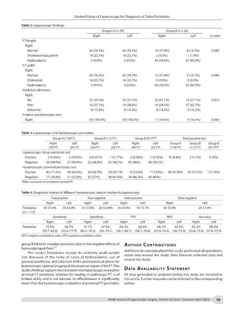
Table 2: Laparoscopic findings
Group A (n = 59) Group B (n = 56)
Right Left Right Left p-value
FT length
Right
Normal 45 (76.3%) 45 (76.3%) 10 (17.9%) 8 (14.3%) 0.000
Shortened but patent 14 (23.7%) 14 (23.7%) 2 (3.6%) 1 (1.8%)
Hydrosalpinx 0 (0.0%) 0 (0.0%) 44 (78.6%) 47 (83.9%)
FT width
Right
Normal 45 (76.3%) 45 (76.3%) 12 (21.4%) 9 (16.1%) 0.000
Distended 14 (23.7%) 14 (23.7%) 0 (0.0%) 0 (0.0%)
Hydrosalpinx 0 (0.0%) 0 (0.0%) 44 (78.6%) 47 (83.9%)
Peritubal adhesions
Right
No 35 (59.3%) 32 (57.1%) 32 (57.1%) 16 (27.1%) 0.972
Fine 16 (27.1%) 16 (28.6%) 16 (28.6%) 37 (62.7%)
Extensive 8 (13.6%) 8 (14.3%) 8 (14.3%) 6 (10.2%)
Positive perchromation test
Right 59 (100.0%) 59 (100.0%) 11 (19.6%) 9 (16.1%) 0.000
Table 3: Laparoscopic and hysteroscopic peristalses
Group A (118 FT) Group B (112 FT) Group B (91 FT) a Total positive test
Right Left Right Left Right Left Group A Group B Group B
(59 FT) (59 FT) (56 FT) (56 FT) (44 FT) (47 FT) (118 FT) (112 FT) (91 FT) a
Laparoscopic distal peristalsis test
Positive 3 (0.05%) 2 (0.03%) 4 (0.07%) 1 (0.17%) 2 (0.04%) 1 (0.02%) 10 (8.4%) 2 (1.7%) 0 (0%)
Negative 56 (94.9%) 57 (96.6%) 52 (46.4%) 55 (98.2%) 42 (46%) 46 (50.5%)
Hysteroscopic proximal peristalsis test
Positive 42 (71.2%) 38 (64.4%) 24 (42.9%) 18 (32.1%) 6 (13.6%) 7 (14.9%) 80 (67.8%) 42 (37.5%) 13 (14%)
Negative 17 (28.8%) 21 (35.6%) 32 (57%) 38 (67.8%) 38 (86.3%) 40 (85%)
a After exclusion of unilateral normal FT
Table 4: Diagnostic indices of different hysteroscopic tests in relation to laparoscopy
True positive True negative False positive False negative
Right Left Right Left Right Left Right Left
Peristalsis 43 37.4% 39 33.9% 32 27.8% 38 33.04% 24 20.9% 18 15.7% 16 13.9% 20 17.4%
(n = 115)
Sensitivity Specificity PPV NPV Accuracy
Right Left Right Left Right Left Right Left Right Left
Peristalsis 72.9% 66.1% 57.1% 67.9% 64.2% 68.4% 66.7% 65.5% 65.2% 66.9%
(59.7–83.6) (52.6–77.9) (43.2–70.3) (54–79.7) (56.1–60.7) (58.7–76.8) (55.4–76.3) (56–73.9) (55.8–73.9) (57.6–75.4)
NPV, negative predictive value; PPV, positive predictive value
group B failed to visualize peristalsis due to the negative effects of Author contrIbutIons
hydrosalpingeal fluid. 23
This study’s limitations include its relatively small sample Atef Darwish conceptualized this study, performed all operations,
size (because of the rarity of cases of hydrosalpinx), use of wrote and revised the study. Dina Darwish collected data and
general anesthesia, and selection of the postmenstrual phase for revised the study.
hysteroscopic optimal imaging of the proximal region of the FT. This
study’s findings support the conclusion that laparoscopic evaluation dAtA AvAIlAbIlIty stAteMent
of distal FT peristalsis, whether for healthy or pathologic FT, is of All data generated or analyzed during this study are included in
limited utility and is not advised. Its effectiveness is significantly this article. Further enquiries can be directed to the corresponding
lower than the hysteroscopic evaluation of proximal FT peristalsis. author.
World Journal of Laparoscopic Surgery, Volume 16 Issue 1 (January–April 2023) 59