
Page 55 - World Journal of Laparoscopic Surgery
P. 55
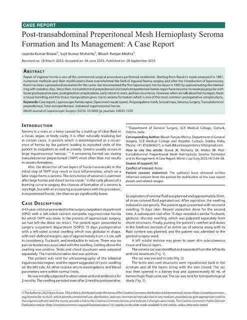
CASE REPORT
            Post-transabdominal Preperitoneal Mesh Hernioplasty Seroma

            Formation and Its Management: A Case Report


                             1
                                                2
            Jayanta Kumar Biswal , Sujit Kumar Mohanty , Bikash Ranjan Mishra 3
            Received on: 18 March 2023; Accepted on: 04 June 2023; Published on: 05 September 2023
             AbstrAct
               Repair of inguinal hernia is one of the commonest surgical procedures performed worldwide. Starting from Bassini’s repair proposed in 1887,
             numerous methods and their modifications have overwhelmed the field of inguinal hernia surgery and after the introduction of laparoscopy
             there has been a procedural revolution for the same. Ger documented the first laparoscopic hernia repair in 1982 by approximating the internal
             ring with stainless clips. Since then, transabdominal preperitoneal and total extraperitoneal hernia repair have become increasingly popular with
             lesser postoperative pain, postoperative complications, early return to work, and less recurrence. However, when we talk about hernia repair, there
             is tissue handling and this tissue manipulation gives rise to seroma formation which is one of the most common postoperative complications.
             Keywords: Case report, Laparoscopic hernia repair, Open mesh repair (open), Polypropylene mesh, Scrotal mass, Seroma, Surgery, Transabdominal
             preperitoneal, Total extraperitoneal, Unilateral inguinoscrotal hernia.
             World Journal of Laparoscopic Surgery (2023): 10.5005/jp-journals-10033-1559


            IntroductIon                                       1–3 Department of General Surgery, SCB Medical College, Cuttack,
            Seroma is a mass or a lump caused by a build-up of clear fluid in   Odisha, India
            a tissue, organ, or body cavity. It is often naturally resolving but   Corresponding Author: Bikash Ranjan Mishra, Department of General
            in certain cases, it persists which is misinterpreted as a recurr-  Surgery, SCB Medical College and Hospital, Cuttack, Odisha, India,
            ence of hernia by the patient leading to repeated visits of the   Phone: +91 8763608377, e-mail: Bikashranjanmishra100@gmail.com
            patient to outpatient as well as anxiety. Seroma usually occurs in   How to cite this article: Biswal JK, Mohanty SK, Mishra BR. Post-
            large inguinoscrotal hernias. 1–4  A remaining hernial sac during   transabdominal  Preperitoneal  Mesh  Hernioplasty  Seroma  Formation
            transabdominal preperitoneal (TAPP) most often than not results   and its Management: A Case Report. World J Lap Surg 2023;16(1):54–56.
            in seroma formation.                               Source of support: Nil
               Also, the dissection of two layers of fascia transversalis in the   Conflict of interest: None
            initial step of TAPP may result in local inflammation, which on a   Patient consent statement:  The author(s) have obtained written
            later stage forms a seroma. The occurrence of seromas is common   informed consent from the patient for publication of the case report
                                           5
            after large hernia and direct hernia repair.  In the early phases of a   details and related images.
            learning curve in surgery, the chances of formation of a seroma is
            very high, but with an increasing acquaintance with the procedure,
            in experienced hands, the chances go significantly lower.
                                                               So aspiration of seromal fluid was planned and approximately 50 mL
                                                               of straw-colored fluid aspirated out. After aspiration, the swelling
            cAse descrIptIon                                   reduced in size greatly. The patient again presented with recurrent
            A 43-year-old man presented to the surgery outpatient department   swelling 15 days later. Repeat aspiration done for the second
            (OPD) with a left-sided indirect complete inguinoscrotal hernia   time. A subsequent visit after 15 days revealed a similar fluctuant,
            for which TAPP was done. In the process of laparoscopic surgery,   globular, discrete swelling, which was palpated separately from
            we had left the distal sac intact. The patient again presented to   chord structures. Finally, putting the patient’s comfort and desire
            surgery outpatient department (SOPD) 15 days postoperative   in the forefront excision of an entire sac of seroma along with its
            with a left-sided scrotal swelling which was globular in shape,   fluid content was planned, and the patient was admitted to the
            with well-defined margins, size of approximately 6 cm × 5 cm, soft   general surgery ward.
            in consistency, fluctuant, and irreducible in nature. There was no   A left scrotal incision was given to open skin subcutaneous
            pain or tenderness associated with the swelling. Getting above the   tissue and fascial layers.
            swelling was positive. Testis and chord structures were palpated   The seroma sac was identified and separated from the left testis
            separately. The transillumination test was positive.  and cod structures (Fig. 1).
               The patient was sent for ultrasonography of the bilateral   The sac was excised in toto (Fig. 2).
            inguinoscrotal region, and the report suggested of cystic swelling   The testis and cord structures were repositioned back in the
            on the left side. All other routine serum investigations and blood   scrotum and all the layers along with the skin closed. The sac
            parameters were within normal limits.              was then opened in a kidney tray and approximately 80 mL of
               He was initially subjected to observation and oral antibiotics for   hemorrhagic fluid came out. The sac was sent for histopathological
            2 months. The swelling persisted even after 2 months postoperative.   study (Fig. 3).


            © The Author(s). 2023 Open Access. This article is distributed under the terms of the Creative Commons Attribution 4.0 International License (https://creativecommons.
            org/licenses/by-nc/4.0/), which permits unrestricted use, distribution, and non-commercial reproduction in any medium, provided you give appropriate credit to
            the original author(s) and the source, provide a link to the Creative Commons license, and indicate if changes were made. The Creative Commons Public Domain
            Dedication waiver (http://creativecommons.org/publicdomain/zero/1.0/) applies to the data made available in this article, unless otherwise stated.