
Page 21 - Journal of WALS
P. 21
WJOLS
Role of Robotic Surgery in the Treatment of Mirizzi Syndrome
Third world association of laparoscopic surgeons conference the rule, robotic retrograde cholecystectomy is an option.
in World Laparoscopy Hospital, DLF Cyber City, Gurgaon, Preoperative ERCP and stenting of the bile duct is usually
Haryana, India (Figs 1 and 2). We also have previously advised. The steps in the surgical procedure in a case of
studied the mechanism and operational ergonomics of the certain diagnosis includes; docking, inserting robotic bipolar
da Vinci surgical robot. References were also made from forceps and hook, dissection of peritoneal adhersions,
available clinical videos. aiming at the right subcostal space, visualization of the
fundus of the GB and GB exposure with careful dissection
RESULTS
of the tissues around the GB, dissection and ligation of the
ERCP and or magnetic resonance cholangiopancreato- cystic artery, retrograde cholecystectomy leading the way
graphy (MRCP) are usually used to define billiary images to the cystic duct, ligature of the cystic duct with stone
anatomically. Results of axial T2-weighted magnetic retrieval and closure of fistula.
resonance imaging (MRI) in a patient having MS and fistula
formation usually show pneumobilia and a suspicion of Port Positions of Robotic Cholecystectomy
fistula. However, the result of the corona T1-weighted image Four ports are used like in conventional laparoscopic
with intravenous gadolinium in same patient usually cholecystectomy with the telescope centered in the umbilical
confirms the presence of such fistulous tract. On the size of
the defect with respect to the common hepatic duct diameter, port (12 mm), one port in the epigastrum (8 mm), two other
results show that in the group of MS where a fistula is 8 mm ports, one midclavicula line below right costal
present; in type 2 the defect is smaller than 33% of the margin and the second a little inferiolateral to it. For the
common hepatic duct diameter, type 3—the defect is 33 to robotic cholecystectomy because of the size of the robot
66% of the diameter of the common hepatic duct and type 4 the working angle is up to 90º and the distance to the target
the defect is 66% of the common hepatic duct diameter. is up to 10 cm (Fig. 3) .
Results also show that nondiagnosis or diagnostic delay DISCUSSION
is usually common, especially in cases where there are no
clinical suspicion and where there are no advanced imaging Treatment of MS depends on the type. In type 1
facilities. It is generally accepted that there is an increased cholecystectomy with choledochostomy to remove the
risk of GB carcinoma in patients with stone disease. From impacted stone is effective. While in type 2 closure of the
the foregoing, particular attention must be focused on the fistula with absorbable material or choledochoplasty with
histology of the cholecystectomy specimen retrieved during the remnant of the GB can be performed. In type 3,
robotic cholecystectomy. Apart from open cholecystectomy choledochoplasty is recommended while type 4 will need a
and laparoscopic-assisted cholecystectomy, purely bilioenteric anastomosis. Robotic surgery is of value in the
laparoscopic cholecystectomy had been done with limited treatment of stage 1 and 2 in combination with preoperative
value in complicated cases of stone disease. Robot-assisted ERCP and intraoperative robotic ultrasound useful in
cholecystectomy has now given way to robotic locating the impacted stone and to partially replicate the
cholecystectomy. In most complicated GB diseases where touch of the surgeons hand which will soon be embedded
multiple peritoneal adhersions and distorted anatomy are as sensors in the newer generation of robots.
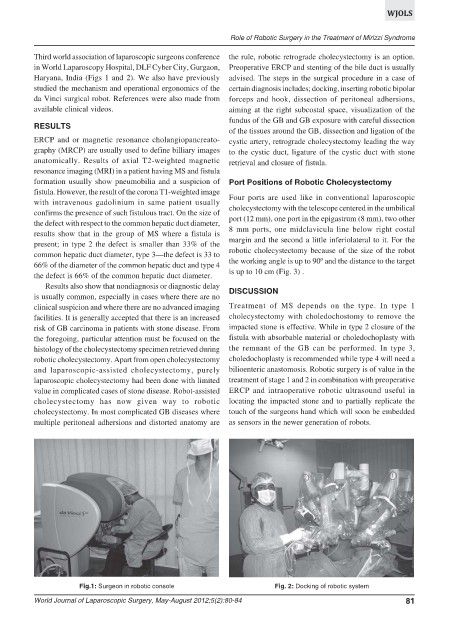
Fig.1: Surgeon in robotic console Fig. 2: Docking of robotic system
World Journal of Laparoscopic Surgery, May-August 2012;5(2):80-84 81