
Page 11 - Journal of Laparoscopic Surgery
P. 11
WJOLS
Laparoscopic Vasectomy vs Laparoscopic Sterilization in Dogs: A Comparison of Two Techniques
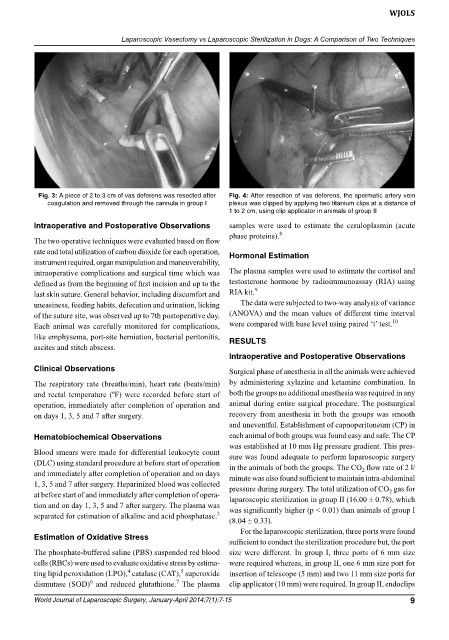
Fig. 3: A piece of 2 to 3 cm of vas deferens was resected after Fig. 4: After resection of vas deferens, the spermatic artery vein
coagulation and removed through the cannula in group I plexus was clipped by applying two titanium clips at a distance of
1 to 2 cm, using clip applicator in animals of group II
Intraoperative and Postoperative observations samples were used to estimate the ceruloplasmin (acute
8
phase proteins).
The two operative techniques were evaluated based on flow
rate and total utilization of carbon dioxide for each operation, hormonal estimation
instrument required, organ manipulation and maneuverability,
intraoperative complications and surgical time which was The plasma samples were used to estimate the cortisol and
defined as from the beginning of first incision and up to the testosterone hormone by radioimmunoassay (RIA) using
last skin suture. General behavior, including discomfort and RIA kit. 9
uneasiness, feeding habits, defecation and urination, licking The data were subjected to two-way analysis of variance
of the suture site, was observed up to 7th postoperative day. (ANOVA) and the mean values of different time interval
10
Each animal was carefully monitored for complications, were compared with base level using paired ‘t’ test.
like emphysema, port-site herniation, bacterial peritonitis, ReSULTS
ascites and stitch abscess.
Intraoperative and Postoperative observations
Clinical observations Surgical phase of anesthesia in all the animals were achieved
The respiratory rate (breaths/min), heart rate (beats/min) by administering xylazine and ketamine combination. In
and rectal temperature (ºF) were recorded before start of both the groups no additional anesthesia was required in any
operation, immediately after completion of operation and animal during entire surgical procedure. The postsurgical
on days 1, 3, 5 and 7 after surgery. recovery from anesthesia in both the groups was smooth
and uneventful. Establishment of capnoperitoneum (CP) in
hematobiochemical observations each animal of both groups was found easy and safe. The CP
was established at 10 mm Hg pressure gradient. This pres-
Blood smears were made for differential leukocyte count sure was found adequate to perform laparoscopic surgery
(DLC) using standard procedure at before start of operation in the animals of both the groups. The CO flow rate of 2 l/
2
and immediately after completion of operation and on days minute was also found sufficient to maintain intra-abdominal
1, 3, 5 and 7 after surgery. Heparinized blood was collected pressure during surgery. The total utilization of CO gas for
2
at before start of and immediately after completion of opera- laparoscopic sterilization in group II (16.00 ± 0.78), which
tion and on day 1, 3, 5 and 7 after surgery. The plasma was was significantly higher (p < 0.01) than animals of group I
3
separated for estimation of alkaline and acid phosphatase.
(8.04 ± 0.33).
For the laparoscopic sterilization, three ports were found
estimation of oxidative Stress
sufficient to conduct the sterilization procedure but, the port
The phosphate-buffered saline (PBS) suspended red blood size were different. In group I, three ports of 6 mm size
cells (RBCs) were used to evaluate oxidative stress by estima- were required whereas, in group II, one 6 mm size port for
5
4
ting lipid peroxidation (LPO), catalase (CAT), superoxide insertion of telescope (5 mm) and two 11 mm size ports for
7
6
dismutase (SOD) and reduced glutathione. The plasma clip applicator (10 mm) were required. In group II, endoclips
World Journal of Laparoscopic Surgery, January-April 2014;7(1):7-15 9