
Page 32 - World Journal of Laparoscopic Surgery
P. 32
Samir S Deolekar et al
inflamed turgid appendix with a normal base (Fig. 3).
There was also a paraovarian cyst present along the left
ovary with minimal free fluid collection in the pouch of
Douglas (Fig. 4). Transposition of the various abdomi-
nal organs was also confirmed (Fig. 5). A laparoscopic
appendectomy with enucleation of paraovarian cyst
was done. An inflamed appendix with inflammatory
infiltrates was confirmed in the histopathology report.
Postoperative recovery was uneventful with patient
discharged on full diet on 4th postoperative day without
any complications.
DISCUSSION
5
Matthew Baillie first demonstrated the complete mirror
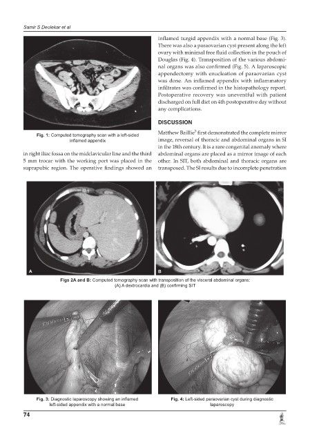
Fig. 1: Computed tomography scan with a left-sided
inflamed appendix image, reversal of thoracic and abdominal organs in SI
in the 18th century. It is a rare congenital anomaly where
in right iliac fossa on the midclavicular line and the third abdominal organs are placed as a mirror image of each
5 mm trocar with the working port was placed in the other. In SIT, both abdominal and thoracic organs are
suprapubic region. The operative findings showed an transposed. The SI results due to incomplete penetration
A B
Figs 2A and B: Computed tomography scan with transposition of the visceral abdominal organs:
(A) A dextrocardia and (B) confirming SIT
Fig. 3: Diagnostic laparoscopy showing an inflamed Fig. 4: Left-sided paraovarian cyst during diagnostic
left-sided appendix with a normal base laparoscopy
74