
Page 29 - World Journal of Laparoscopic Surgery
P. 29
WJOLS
Osseous Metaplasia of Endometrium: A Very Rare Entity
A B
C
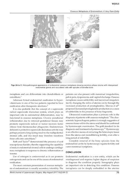
Figs 2A to C: Histopathological appearance of endometrial osseous metaplasia showing secretory phase stroma with interspersed
endometrial glands and vacuolated cells with spicules of lamellar bone
metaplasia and can differentiate into chondroblasts or patients can also present with menstrual irregularities,
osteoblasts. 1 pelvic pain, dyspareunia, and vaginal discharge. Osseous
Adamson linked endometrial ossification to hyper- metaplasia causes subfertility and menstrual irregulari-
vitaminosis in one of his two patients reported to have ties by changing the milieu of uterine cavity through the
11
ossification after therapeutic abortions. 2 increased production of prostaglandins. Marcus et al
It is also probable that the concept of a superoxide proposed increased prostaglandin production as a cause
radical superoxide dismutase system, which plays an of subfertility in the presence of bony fragments.
important role in endometrial differentiation, may be Ultrasound examination plays a primary role in the
7
functional in osseous metaplasia. Chronic postabortal diagnosis of patients with osseous metaplasia. The char-
inflammation due to retained gestational tissues may acteristic hyperechogenic pattern is strongly suggestive of
promote superoxide radical or tumor necrosis factor osseous tissue within the uterus and should be confirmed
release from thermonuclear phagocytes. Endometrium by hysteroscopic examination. The gold standard for its
12
deficient in protective superoxide dismutase activity may diagnosis and treatment is hysteroscopy. Hysteroscopy
perhaps present a long-lasting insult to the multipotential is an effective means of extracting the heterotopic tissue
stromal cells, and this insult may therefore transform from the uterus and reestablishing fertility, even after a
these cells into osteoblasts. 9 long period of infertility.
10
Roth and Taylor demonstrated the presence of acid Complete removal of the bony spicules from the
mucopolysaccharides, thereby supporting the capability endometrial cavity by hysteroscopy regains fertility and
of mature endometrial stromal cells to undergo cartilagi- cures menstrual symptoms.
nous metaplasia in response to chronic inflammation or
trauma. CONCLUSION
Use of estrogen is controversial as it can promote Endometrial ossification is a rare entity, which can be
osteogenesis and can be one of the causes of endometrial misdiagnosed and requires higher degree of suspicion
ossification. to diagnose the condition properly. Sonography plays
The most common presentation of osseous metapla- an important role in detecting this condition. Osseous
sia of endometrium is usually secondary infertility. The metaplasia can be deeply embedded in the uterine
World Journal of Laparoscopic Surgery, May-August 2017;10(2):69-72 71