
Page 35 - World Journal of Laparoscopic Surgery
P. 35
WJOLS
WJOL S
10.5005/jp-journals-10033-1309
Xanthogranulomatous Cholecystitis
CASE REPORT
Xanthogranulomatous Cholecystitis
2
1 Hana Alhomoud, Mohamed Abdelmohsen
ABSTRACT biliary radicles, possibility of cholangiocarcinoma as
Xanthogranulomatous cholecystitis is a rare, benign, chronic described by sonarist and suggestion of cholecystitis
inflammatory disease of the gallbladder (GB). Its importance lies with fluid collection in CT conclusion. The patient had
in the fact that imaging studies and intraoperative appearance endoscopic retrograde cholangiopancreatography with
may mimic tumor of the GB. Xanthogranulomatous cholecystitis papillotomy and sweeping to the CBD with balloon
is difficult to diagnose pre- or intraoperatively and remains a
challenge in medical practice. The definitive diagnosis depends catheter, with small amount of pus coming from the GB
on the histopathologic examination. as described by the interventional radiologist.
Laparoscopic cholecystectomy was started, which
Keywords: Gallbladder cancer, Surgery, Xanthogranulomatous
cholecystitis. was converted to open cholecystectomy. The GB wall
was thickened and the serosa was surrounded by dense
How to cite this article: Alhomoud H, Abdelmohsen M. Xantho-
granulomatous Cholecystitis. World J Lap Surg 2017;10(2):77-79. fibrous adhesions, which were attached to adjacent
hepatic parenchyma and transverse colon. There was a
Source of support: Nil
small-sized abscess in the GB wall. Dissection between
Conflict of interest: None the GB serosa and hepatic parenchyma was difficult
leading to subtotal cholecystectomy. Cross-section
INTRODUCTION through the wall revealed multiple yellow-colored,
nodule-like lesions, and there were also multiple black-
Xanthogranulomatous cholecystitis (XGC) is an uncom- pigmented gallstones.
mon variant of chronic cholecystitis characterized by The pathologic findings showed the collections of
the presence of grayish yellow nodules or streaks in foamy histiocytes containing abundant lipid in the cyto-
the gallbladder (GB) wall, mainly caused by lipid-laden plasm and admixed lymphoid cells. Histologically, it was
1
macrophages. Although well-defined pathologically, confirmed as XGC.
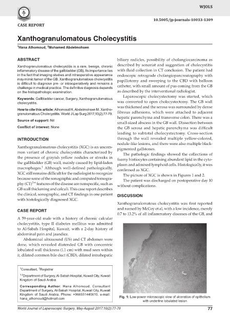
XGC still remains difficult for the radiologist to recognize The picture of XGC is shown in Figures 1 and 2.
because some of the sonographic and computed tomogra- The patient was discharged on postoperative day 10
2-4
phy (CT) features of the disease are nonspecific, such as without complications.
GB wall thickening and calculi. This case report describes
the clinical, sonographic, and CT findings in one patient DISCUSSION
with histologically diagnosed XGC.
Xanthogranulomatous cholecystitis was first reported
CASE REPORT and named by McCoy et al, with a low incidence, merely
0.7 to 13.2% of all inflammatory diseases of the GB, and
A 59-year-old male with a history of chronic calcular
cholecystitis, type II diabetes mellitus was admitted
to Al-Sabah Hospital, Kuwait, with a 2-day history of
abdominal pain and jaundice.
Abdominal ultrasound (US) and CT abdomen were
done, which revealed distended GB with concentric
lobulated wall thickness (1.1 cm) with mud seen within
it, dilated common bile duct (CBD), dilated intrahepatic
1 Consultant, Registrar
2
1,2 Department of Surgery, Al-Sabah Hospital, Kuwait City, Kuwait
Kingdom of Saudi Arabia
Corresponding Author: Hana Alhomoud, Consultant
Department of Surgery, Al-Sabah Hospital, Kuwait City, Kuwait
Kingdom of Saudi Arabia, Phone: +966551440610, e-mail:
hana_alhomoud@hotmail.com Fig. 1: Low-power microscopic view of ulceration of epithelium
with underline lobulated lesion
World Journal of Laparoscopic Surgery, May-August 2017;10(2):77-79 77