
Page 17 - Textbook of Practical Laparoscopic Surgery by Dr. R. K. Mishra
P. 17
16 SECTION 1: Essentials of Laparoscopy
■ ■The incidence of injury to adhesion although not Veress needle abdominal insufflation or secondary port
eliminated is significantly reduced by entry into the placement after pneumoperitoneum has already been
peritoneal cavity under direct vision. established.
■ ■There is a decreased risk of injury to the retroperitoneal
vessels. The trocar is blunt, and the angle of entry allows Open Fielding Technique
the surgeon to maneuver the cannula at an angle, This technique developed by Fielding in 1992 involves
which avoids viscera, while still assuring peritoneal a small incision over the everted umbilicus at a point
placement. where the skin and peritoneum are adjacent. Fielding
■ ■The risk of extraperitoneal insufflation is eliminated. technique is useful in patients with abdominal incisions
Placement under direct vision ensures that insufflation from previous surgery provided there is no midline
of gas is actually into the peritoneal cavity. incision, portal hypertension and recanalized umbilical
■ ■The likelihood of hernia formation is decreased vein, and umbilical abnormalities, such as urachal cyst,
because the fascia is closed as part of the technique. sinus or umbilical hernia. Thorough skin preparation of
■ ■Increasing number of surgeons performing laparoscopy the umbilicus is carried out and the everted umbilicus
without experience and in these group open technique is incised from the apex in a caudal direction. Two small
may be easy. retractors are inserted to expose the cylindrical umbilical
■ ■Useful in muscular man and children with strong tube running from the undersurface of the umbilical skin
abdominal wall. down to the linea alba.
■ ■Useful for gynecologists or surgeon lacking enough This tube is then cut from its apex downward
upper arm strength to elevate the abdominal wall of toward its junction with the linea alba. Further, blunt
patient. dissection through this plane permits direct entry into
■ ■An open technique, which involves creating a the peritoneum. Once the peritoneal cavity is breached,
minilaparotomy into which a special cannula is the primary port can be inserted directly, and insufflation
inserted, may be adopted. This procedure has its own started. A blunt internal trocar facilitates insertion of this
complications and requires skilled execution. port and an external grip that can be attached to the port
assist to secure it in position. Suture is usually not required
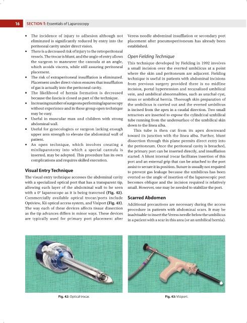
Visual Entry Technique to prevent gas leakage because the umbilicus has been
The visual entry technique accesses the abdominal cavity everted so the angle of insertion of the laparoscopic port
with a specialized optical port that has a transparent tip, becomes oblique and the incision required is relatively
allowing each layer of the abdominal wall to be seen small. However, one may be needed to stabilize the port.
with a 0° laparoscope as it is being traversed (Fig. 42).
Commercially available optical trocar/ports include Scarred Abdomen
Optiview, Kii optical access system, and Visiport (Fig. 43). Additional precautions are necessary during the access
The way each of these devices affects tissue dissection procedure in patients with abdominal scars. It may be
as the tip advances differs in minor ways. These devices inadvisable to insert the Veress needle below the umbilicus
are typically used for primary port placement after in a patient with a scar in this area (or an umbilical hernia).
Fig. 42: Optical trocar. Fig. 43: Visiport.