
Page 47 - Journal of World Association of Laparoscopic Surgeons
P. 47
CASE REPORT
Laparoscopic Management of Median Arcuate Ligament
Syndrome: Single Center Experience
3
2
1
Eppa Vimalakar Reddy , Gourang Shroff , Vemula Bala Reddy , Akella V Phanendra Somayajulu 4
AbstrAct
Median arcuate ligament syndrome (MALS) is a rare disease caused as a result of extrinsic compression by diaphragmatic fibers arching on
the celiac artery at its point of origin from the abdominal aorta. Patients suffering from MALS presented with weight loss, nausea, vomiting,
and postprandial epigastric pain. Often misdiagnosed with dyspepsia or acid peptic disease, this syndrome is a diagnosis by exclusion, after
excluding commoner causes of the upper abdomen pain. It is diagnosed with computed tomographic (CT) angiography and treated with
various modalities, including laparoscopic or open division of fibers of MAL, which cause extrinsic pressure. We report a series of three cases of
MALS diagnosed and managed at our center, using laparoscopic division of the fibers and release of the celiac artery.
Keywords: Celiac artery compression syndrome, Dunbar syndrome, Laparoscopy, Median arcuate ligament syndrome, Minimal invasive.
World Journal of Laparoscopic Surgery (2019): 10.5005/jp-journals-10033-1358
IntroductIon 1–4 Department of Surgical Gastroenterology, Sunshine Hospitals,
The median arcuate ligament (MAL) is an arch of diaphragmatic Secunderabad, Telangana, India
fibers crossing the aorta, superior to the celiac artery origin and at Corresponding Author: Eppa Vimalakar Reddy, Department of
1–3
the level of diaphragmatic insertion. Its lower insertion crosses Surgical Gastroenterology, Sunshine Hospitals, Secunderabad,
1–5
the proximal part of the celiac artery. Telangana, India, Phone: +91 9573201103, e-mail: vimalakarreddy@
MALS is a rare disease caused by the extrinsic compression on gmail.com
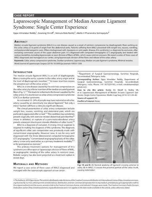
the celiac artery by inferior insertion of the median arcuate ligament How to cite this article: Reddy EV, Shroff G, Reddy VB,
1–5
fibers (Fig. 1). This leads to ischemia to the bowel supplied by the et al. Laparoscopic Management of Median Arcuate Ligament Syn-
celiac artery. It is also known as celiac artery compression syndrome drome: Single Center Experience. World J Lap Surg 2019;12(1):39–42.
(CACS) or dunbar syndrome. Source of support: Nil
An estimated 10–24% of people may have indentation of celiac Conflict of interest: None
6
artery caused by an abnormally low placed ligament. But only a
minor fraction will have a clinically significant disease.
The clinical presentation of celiac artery compression include
weight loss, nausea, vomiting, and abdominal pain, which are
3–5
particularly aggravated after a meal. The condition may sometimes
12
present atypically with exercise-related abdominal pain/diarrhea
(more in athletes), or rupture of a pancreaticoduodenal artery
7,8
pseudo-aneurysm (due to post-stenotic dilatation of celiac trunk).
MALS is a diagnosis of exclusion. A strong clinical suspicion is
required in making the diagnosis of this syndrome. The diagnosis
of significant celiac axis compression was previously made with
conventional angiography. However now, it can be very well
diagnosed with the three-dimensional computed tomographic
2
(CT) angiography. Convensional angiography/plasty of the celiac
9,10
artery is now only used either as a primary treatment modality
11
or for postoperative stenosis.
The primary treatment options for management of this
syndrome are either open or laparoscopic division of fibers of MAL,
or angiographic stenting of the celiac artery in resistant cases.
Robotic surgery has also been projected as a treatment option in
recent days.
MAterIAls And Methods Figs 1A and B: (A) Normal anatomy of ligament crossing anterior to
We report a case series of three cases of MALS diagnosed and aorta; (B) In MALS, it crosses the proximal portion of the celiac trunk,
managed with the laparoscopic approach at our center. causing indentation
© The Author(s). 2019 Open Access This article is distributed under the terms of the Creative Commons Attribution 4.0 International License (https://creativecommons.
org/licenses/by-nc/4.0/), which permits unrestricted use, distribution, and non-commercial reproduction in any medium, provided you give appropriate credit to
the original author(s) and the source, provide a link to the Creative Commons license, and indicate if changes were made. The Creative Commons Public Domain
Dedication waiver (http://creativecommons.org/publicdomain/zero/1.0/) applies to the data made available in this article, unless otherwise stated.