Page 9 - World Journal of Laparoscopic Surgery
P. 9

Case Report Related to Laparoscopic Cholecystectomy

            PATHOLOGIC FINDINGS

            1. Nodules with nonpolarizing yellow bile pigment material/
               precipitate and cholesterol crystals associated with foreign
               body (Figs 2 to 4) type chronic granulomatous inflammation
               on serosa of appendix and omentum. No evidence of
               carcinoma.
            2. Mild periappendicitis. The lumen is slightly distended with
               hemorrhagic fluid.

            COMMENT

            A review of the history of minimal access surgery by
                       1
            Dr RK Mishra  goes back to 1585. Laparoscopic surgery was
            originally popular amongst gynecologists and orthopedic
            surgeons. The first scientifically documented laparoscopic
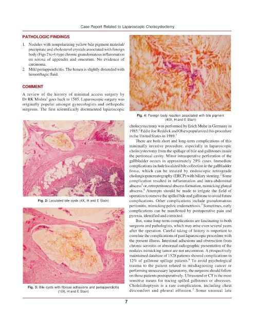
                                                                   Fig. 4: Foreign body reaction associated with bile pigment
                                                                                 (40X, H and E Stain)
                                                               cholecystectomy was performed by Erich Muhe in Germany in
                                                                    1
                                                               1985.  Eddie Joe Reddick and Olsen popularized this procedure
                                                               in the United States in 1989. 1
                                                                  There are both short and long-term complications of this
                                                               minimally invasive procedure, especially in laparoscopic
                                                               cholecystectomy from the spillage of bile and gallstones inside
                                                               the peritoneal cavity. Minor intraoperative perforation of the
                                                               gallbladder occurs in approximately 29% cases. Immediate
                                                               complications include localized bile collection in the gallbladder
                                                               fossa, which can be treated by endoscopic retrograde
                                                                                                           2
                                                               cholangiopancreatography (ERCP) with biliary stenting.  Some
                                                               complication resulted in inflammation and intra-abdominal
                                                                     3
                                                               abscess  or, retroperitoneal abscess formation, mimicking gluteal
                                                                      4
                                                               abscess.  Attempts should be made to irrigate the field of
                                                               operation to remove the spilled bile and gallstone to avoid future
                   Fig. 2: Loculated bile cysts (4X, H and E Stain)  complications. Other complications include granulomatous
                                                                                                  5
                                                               peritonitis, mimicking pelvic endometriosis.  Sometimes, early
                                                               complications can be manifested by postoperative pain and
                                                               pyrexia, identified and corrected.
                                                                  But, some long-term complications are fascinating to both
                                                               surgeons and pathologists, which may arise even several years
                                                               after the operation. Careful taking of history is important to
                                                               correlate the complications of past laparoscopic procedure with
                                                               the present illness. Intestinal adhesions and obstruction from
                                                               chronic serositis or abnormal radiographic presentation of the
                                                               nodules mimicking tumor are not uncommon. A prospectively
                                                               maintained database of 1528 patients showed complications in
                                                                                            6
                                                               12% of gallstone spillage patients.  To avoid psychological
                                                               trauma to the patient related to misdiagnosing cancer or
                                                               performing unnecessary laparotomy, the surgeons should follow
                                                               on those patients postoperatively. Ultrasound or CT is the most
                                                               sensitive means for tracing spilled gallstones or abscesses.
                                                               Cholelithoptysis is a rare complication, including chest
              Fig. 3: Bile cysts with fibrous adhesions and periappendicitis                  7
                             (10X, H and E Stain)              discomfort and pleural effusion.  Some unusual late

                                                             7
   4   5   6   7   8   9   10   11   12   13   14