
Page 17 - WALS Journal
P. 17
10.5005/jp-journals-10033-1170
Abhiijit Sharadchandra Joshi
CASE REPORT
Laparoscopic Management of Renal Hydatid Cyst
Abhiijit Sharadchandra Joshi
ABSTRACT establishing pneumoperitoneum with the closed method,
I submit herewith, a case report of a 55-year-old male farmer, using Veress’ needle and CO insufflation, the trocars were
2
who developed a large left renal lower pole hydatid cyst. He inserted. Two 10 mm and two 5 mm trocars were used.
was successfully treated laparoscopically in April 2007, via the Dissection was commenced (Fig. 3) by reflecting the
transperitoneal access. There were no intraoperative
complications and over a 2.5 years follow-up period. He was descending colon medially after incising the lateral
essentially asymptomatic and disease free. To the best of my peritoneal fold so as to enter the retroperitoneal space. The
knowledge, this is only the fourth reported case of laparoscopic cyst wall was well demarcated. The cyst was then
treatment of renal hydatid cyst.
surrounded from all sides by hypertonic saline-soaked gauze
Keywords: Renal hydatid, Laparoscopically transperitoneal. pieces to avoid contamination of the peritoneal cavity in
How to cite this article: Joshi AS. Laparoscopic Management the event of spillage of the contents of the cyst. The second
of Renal Hydatid Cyst. World J Lap Surg 2012;5(3):150-152. 10 mm trocar was then introduced under laparoscopic vision
Source of support: Nil directly into the cyst (Fig. 4). No spillage occurred at the
trocar entry site during or after the entry. A 10 mm suction
Conflict of interest: None declared
cannula was then inserted into the cyst and the contents
INTRODUCTION were sucked out (Fig. 5). Hypertonic saline was then instilled
into the cyst through the second channel on the suction
Hydatid disease is endemic in cattle and sheep-raising
regions of the world. The treatment of hydatid cysts is cannula, was kept in situ for 10 minutes and was then sucked
principally surgical. With advances in laparoscopic out. Then the laparoscope was passed into the cyst to directly
techniques and equipment, hydatid disease has become visualize and confirm complete evacuation (Fig. 6). After
manageable by the same. this the scope was reinserted through the subumbilical
10 mm trocar and the intracystic 10 mm trocar was
CASE REPORT withdrawn out of the cyst. A cystotomy was then performed
A 55-year-old farmer presented to our hospital in March to gain access into the cyst after which the endocyst was
2007 with left-sided abdominal pain and lump in left side removed in toto and placed in endo bag. The remnant
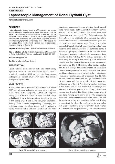
of abdomen. Ultrasonography (USG) and computed ectocyst was deroofed (Fig. 7) at multiple places where it
tomographic (CT) scan of the abdomen revealed a large was bare, taking care not to injure the descending
hydatid cyst, 15 cm in diameter, arising from the lower pole mesocolon. These chunks of ectocyst were all extracted with
of left kidney (Figs 1 and 2). He was given albendazole the endocyst using the endobag. After confirming
600 mg OD for 2 weeks preoperatively. The surgery was hemostasis at the edges, the resulting cavity was packed
performed under general anesthesia, with the patient in with greater omentum held in position with 4-5 silk stitches.
supine position with a left side elevation of 15º. After A 28 Fr tube drain was passed through the lateral trocar site
Fig. 1: CT image of LT renal hydatid 1 Fig. 2: CT image of LT renal hydatid 2
150
JAYPEE