
Page 46 - Journal of Laparoscopic Surgery
P. 46
Mohammed Arifuzaman, Asna Samreen
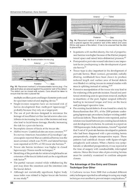
Fig. 14: Placement method 1 of bioabsorbable hernia plug–The
disk is placed against the posterior wall of the defect and tubes
fill the void space of the defect. It has to be ensured that the disk
is placed flat
• In patients with morbid obesity, the risk of preperito-
neal hernias was higher because of the thicker preperi-
toneal space and raised intra-abdominal pressure. 42
Fig. 13: Bioabsorbable hernia plug
• Postoperative port-site wound infection is one impor-
tant factor predisposing to the development of port-
site hernia. 43
• Trocar type is also important in the development of
port-site hernia. Blunt (conical, pyramidal, radially
dilating, nonbladed) have been shown to produce
reduced length and surface area of fascial defects
over bladed or cutting trocars in animal studies with
Fig. 15: Placement method 2 of bioabsorbable hernia plug –The muscle splitting instead of cutting. 44,45
disk and tubes are placed against the posterior wall of the defect. • Extensive manipulation of the trocar site may lead to
The defect can be closed with sutures. Care should be taken to
ensure that the disk is placed flat the widening of the port-site incision. Fascial and peri-
toneal stretching seen in specimen removal, multiple
multiple ancillary ports and larger diameter ports used re-insertions of the port, higher surgical difficulty
for specimen removal and stapling device. 34 leading to increased torque and force on the fascia
• Single-incision surgeries have an increased risk of and prolonged operative time.
35
hernia development than multi-port laparoscopy • Pre-existing fascial defects–It was found in a study by
probably because they rely on a larger port.
• The use of port devices designed to minimize the Ramachandran that 18%, of the 2100 patients under-
going laparoscopic procedures, had pre-existing umbil-
leakage of insufflated air like fascial screws also con- ical fascial defects. These defects were repaired, and no
tributes in increasing the size of the incision and may relation was found between pre-existing fascial defects
also lead to facial tissue damage, thereby increasing
the risk for a port-site hernia. and development of a hernia. In contrast, in a report on
46
• Incomplete closure of fascia at the trocar site. 1300 laparoscopic cholecystectomies, Azurin reported
• Midline trocars: Uumbilical sites are more common. 36,37 that 9 out of 10 port-site hernias developed in patients
In a survey American Association of Gynecologic Lap- who had been diagnosed with a pre-existing hernia
aroscopists reported that an umbilical hernia was the preoperatively, despite intraoperative repair. These
most common which was 75.70% and lateral hernias patients had umbilical closure with figure-of-eight
were reported at 23.70% of 152 trocar site hernias. 38 polyglycolic acid sutures. When a hernia was symp-
• Trocar site hernia incidence was higher in closed tomatic or identified preoperatively, it was repaired at
laparoscopy (Veress needle technique). 39 the time of surgery with nonabsorbable, interrupted
• Stretching of the port-site for retrieval might lead to an sutures. Hence the trocar sites of pre-existing hernias
extension of the fascial defect and can be a significant must be carefully examined to confirm adequate
risk factor. 40 closure.
• The partial vacuum created while withdrawing the The Advantage of One Entry and Closure
port may draw the omentum and the intestines into
the fascial defect. Technique Over Other
• Although not statistically significant, higher body A Cochrane review from 2008 that evaluated different
mass index was related to higher trocar site hernias entry techniques reported no advantage in using any single
47
in one study. 41 technique over another to prevent major complications.
98