
Page 34 - Journal of Laparoscopic Surgery
P. 34
Jitendra Kumar, Rajni Raina
1 January 2012–1 January 2016 has been screened. Total of abdominal wall as this helps in reducing the dead space
thirteen cases (n = 13) of a non-midline hernia found to and preventing the postoperative seroma formation.
be eligible for study in term of detail availability of case After removal of fatty deposits around the defect and
record. Apart from demographic and clinical profile, e.g., thorough hemostasis, we measure defect size to plan the
age, sex, weight, symptoms and its duration, comorbidity, placement of adequate size of mesh. As per recommenda-
past history of surgery; total duration of surgery (from tion mesh should be of a size which can overlap beyond 5
9
making first incision to taking last suture), all intra and cm. of defect margin. In cases where mesh had to place
postoperative events, e.g., size and location of defect, over the defect situated near the iliac or pubic bone in
intra and postoperative different complications, follow the lower part of abdomen we reflected the peritoneum
up period and reported recurrence looked in to and after dissecting it and mesh been tacked over ligament
evaluated in detail. or in some case over bone. In upper abdominal hernia
defect, we dissect the falciform ligament to place the
Surgical Techniques mesh in subcostal region properly. In our all cases we
used composite (coated polypropylene, proceed) mesh.
All cases were done as an elective case at Lady Hardinge
Medical College and Smt. S.K. Hospital, New Delhi. For During defect size measurement and fixation process
preoperative preparation, all patients were made medi- of mesh, as per recommendation, we reduce the intra-
cally fit in term of any associated medical comorbidities, abdominal CO pressure to 5–7 mm of Hg. We fixed
2
diabetes control, cessation of smoking, weight reduction, the mesh with four quadrants trans fascial suture and
etc. Operative techniques followed according to SAGES circumferentially double crowning with non-absorbable
10
guideline and adhered to standard protocols based on titanium tack (Protack, Covidien). Again after being
6-8
different recommended trials. All cases were done assured about hemostasis and other intra-abdominal
under general anesthesia. All cases involving hernia findings, we remove the trocars under vision and suture
below umbilical line had been routinely catheterized after the 10 mm port site with port closure needle in two-layer
induction of anesthesia and catheter was removed soon while rest of the port been closed with only one layer
after completion of surgery. Strict antiseptic and aseptic of skin closure. We usually place large cotton ball com-
protocols have been followed. pression elastic pressure dressing over the large defect
The procedure starts with the creation of pneumoperi- thinking to reduce postoperative seroma. Postoperatively
toneum by a close technique using veres needle mostly at for inspection of port site wound and hernial site, we
palmer point or infra/supra umbilical location depending removed the dressing of the wound after 48 hours.
on the location of a hernia. First port inserted blindly and
rest of the port under camera vision. In all cases, three RESULTS
ports, one camera 10–11 mm and two working port of During four years, 13 cases of nonmidline abdominal
5 mm has been used. Placement of ports depends on site wall hernia found to be operated by the main author.
of a hernia. Mostly port has been placed on the lateral Out of thirteen cases, ten (76.92%) were female, and three
side of the abdomen with camera port in the center and (23.07%) were male with their mean age of 43 +/– 9.30
9
at the possible distant location from defect area. After years (SD = 11.41). The range for age were 24–64 years.
a thorough inspection of inside the abdomen first thing Mean weight of the patients were 72.846 kg (SD = 13.369)
we do is adhesiolysis using electrocautery or a harmonic in range of 52–98 kg (Table 1).
scalpel. A lot of patience and precautions are required for The average duration of hospital stay for the patients
this step to prevent complication like bowel injury. Then were 6.61 days (SD = 4.17) in range of 3–19 days. Average
reduction of the abdominal contents from the hernia follow-up periods were 21.15 months (SD = 11.857) in
sac performed gently and carefully. We do not close the range of 1–40 months (Table 1)
defect or approximate its margin by any means, rather we All patients were having a common complaint of
pull the redundant sac and tack this to adjoining normal swelling, with five patients (38.46%) having pain along
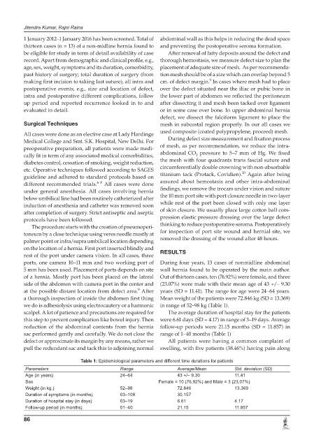
Table 1: Epidemiological parameters and different time durations for patients
Parameters Range Average/Mean Std. deviation (SD)
Age (in years) 24–64 43 +/– 9.30 11.41
Sex Female = 10 (76.92%) and Male = 3 (23.07%)
Weight (in kg.) 52–98 72.846 13.369
Duration of symptoms (in months) 03–108 30.157
Duration of hospital stay (in days) 03–19 6.61 4.17
Follow-up period (in months) 01–40 21.15 11.857
86