
Page 61 - World Journal of Laparoscopic Surgery
P. 61
Congenital Midgut Malrotation Presenting as Acute Duodenal Obstruction in an Adult—Laparoscopic Management
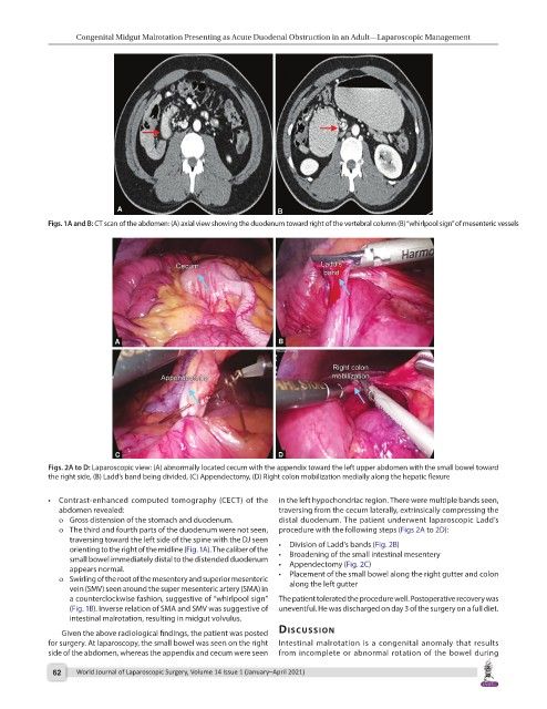
Figs. 1A and B: CT scan of the abdomen: (A) axial view showing the duodenum toward right of the vertebral column (B) “whirlpool sign” of mesenteric vessels
Figs. 2A to D: Laparoscopic view: (A) abnormally located cecum with the appendix toward the left upper abdomen with the small bowel toward
the right side, (B) Ladd’s band being divided, (C) Appendectomy, (D) Right colon mobilization medially along the hepatic flexure
• Contrast-enhanced computed tomography (CECT) of the in the left hypochondriac region. There were multiple bands seen,
abdomen revealed: traversing from the cecum laterally, extrinsically compressing the
o Gross distension of the stomach and duodenum. distal duodenum. The patient underwent laparoscopic Ladd’s
o The third and fourth parts of the duodenum were not seen, procedure with the following steps (Figs 2A to 2D):
traversing toward the left side of the spine with the DJ seen
orienting to the right of the midline (Fig. 1A). The caliber of the • Division of Ladd’s bands (Fig. 2B)
small bowel immediately distal to the distended duodenum • Broadening of the small intestinal mesentery
appears normal. • Appendectomy (Fig. 2C)
o Swirling of the root of the mesentery and superior mesenteric • Placement of the small bowel along the right gutter and colon
vein (SMV) seen around the super mesenteric artery (SMA) in along the left gutter
a counterclockwise fashion, suggestive of “whirlpool sign” The patient tolerated the procedure well. Postoperative recovery was
(Fig. 1B). Inverse relation of SMA and SMV was suggestive of uneventful. He was discharged on day 3 of the surgery on a full diet.
intestinal malrotation, resulting in midgut volvulus.
Given the above radiological findings, the patient was posted dIscussIon
for surgery. At laparoscopy, the small bowel was seen on the right Intestinal malrotation is a congenital anomaly that results
side of the abdomen, whereas the appendix and cecum were seen from incomplete or abnormal rotation of the bowel during
62 World Journal of Laparoscopic Surgery, Volume 14 Issue 1 (January–April 2021)