
Page 57 - World Journal of Laparoscopic Surgery
P. 57
CASE REPORT
Role of Laparoscopy in Gastric Trichobezoar: A Case Report
and Review of Laparoscopic Techniques in Pediatric and
Adolescents
1
2
Prashant Jain , Ashish Prasad , Sarika Jain 3
AbstrAct
This article presents a case report of the laparoscopic removal of a large gastric trichobezoar in a 13-year-old girl. We reviewed the various
laparoscopic techniques and their modifications described in the literature for removal of gastric trichobezoar. Advantages and disadvantages
of various techniques were also discussed.
Keywords: Children, Gastric trichobezoar, Laparoscopy.
World Journal of Laparoscopic Surgery (2021): 10.5005/jp-journals-10033-1438
IntroductIon 1,2 Department of Pediatric Surgery and Pediatric Urology, BLK
Trichobezoar (ball of hair) is accumulation of hair in stomach Superspeciality Hospital, New Delhi, India
and small intestine (Rapunzel syndrome). It is a rare condition 3 Department of Radiology, Doda Imaging, New Delhi, India
1
usually seen in adolescent girls with a psychiatric disorder. Corresponding Author: Prashant Jain, Department of Pediatric
The management of gastric trichobezoar includes endoscopic/ Surgery and Pediatric Urology, BLK Superspeciality Hospital, New
surgical removal along with the treatment of psychiatric instability. Delhi, India, Phone: +91 9582413828, e-mail: docpedsurg@gmail.com
Various techniques have been used which includes laparotomy, How to cite this article: Jain P, Prasad A, Jain S. Role of Laparoscopy
endoscopy, laparoscopy, and laser fragmentation. We report a case in Gastric Trichobezoar: A Case Report and Review of Laparoscopic
of laparoscopic removal of a large trichobezoar in a 13-year-old girl Techniques in Pediatric and Adolescents. World J Lap Surg 2021;
and reviewed various laparoscopic techniques and its modifications 14(1):58–60.
described for removal of gastric trichobezoar. Source of support: Nil
Conflict of interest: None
cAse report
A 13-year-old female, presented with recurrent abdominal
pain and vomiting, which had increased in severity for the last saline wash was given. The trichobezoar was retrieved piecemeal
2 days. Initial evaluation with ultrasound abdomen was normal. with minimal fragmentation through the umbilical port (Fig. 2).
In view of persistent pain and fullness of the upper abdomen, The procedure took about 2 hours and 30 minutes. The size of the
she was evaluated by a gastroenterologist. The child underwent bezoar was 12 × 10 × 7 cm weighing about 200 gm.
upper gastrointestinal endoscopy which revealed a large
trichobezoar involving the stomach and extending into the whole
of the duodenum and proximal jejunum. Endoscopic removal was
attempted twice but only the tail part could be removed. The girl
was then planned for laparoscopic removal of the trichobezoar.
A 12 mm infraumbilical port was used and two 5 mm ports
in epigastrium and left hypochondrium. Pneumoperitoneum
was created using 12 mm pressure. Gastrotomy incision of about
6 cm was made over anterior wall of the stomach. To stabilize
the stomach, two stay sutures were taken through the edge of
the stomach wall and were brought out through the anterior
abdominal wall. With the help of graspers and suction, the bezoar
was gradually separated avoiding any peritoneal contamination.
Our job was made easier by previous endoscopic mobilization of
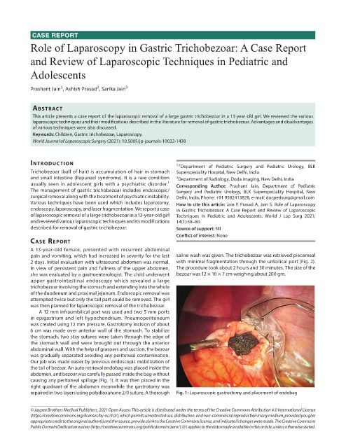
the tail of bezoar. An auto retrieval endobag was placed inside the
abdomen, and bezoar was carefully passed inside the bag without
causing any peritoneal spillage (Fig. 1). It was then placed in the
right quadrant of the abdomen meanwhile the gastrotomy was
repaired in two layers using polydioxanone 2/0 suture. A thorough Fig. 1: Laparoscopic gastrostomy and placement of endobag
© Jaypee Brothers Medical Publishers. 2021 Open Access This article is distributed under the terms of the Creative Commons Attribution 4.0 International License
(https://creativecommons.org/licenses/by-nc/4.0/), which permits unrestricted use, distribution, and non-commercial reproduction in any medium, provided you give
appropriate credit to the original author(s) and the source, provide a link to the Creative Commons license, and indicate if changes were made. The Creative Commons
Public Domain Dedication waiver (http://creativecommons.org/publicdomain/zero/1.0/) applies to the data made available in this article, unless otherwise stated.