
Page 65 - World Journal of Laparoscopic Surgery
P. 65
A Novel Technique Using Mesh to Repair a Recurrent Large Indirect Inguinoscrotal Hernia
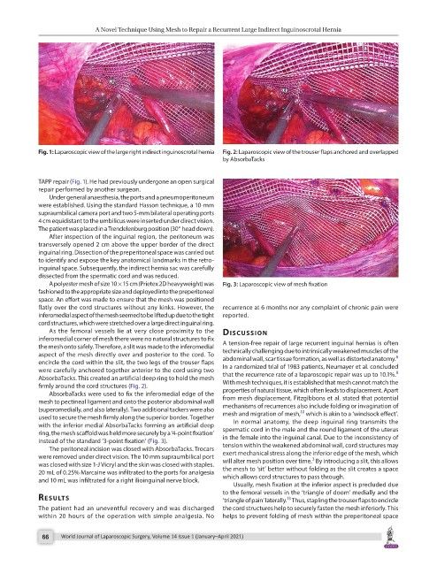
Fig. 1: Laparoscopic view of the large right indirect inguinoscrotal hernia Fig. 2: Laparoscopic view of the trouser flaps anchored and overlapped
by AbsorbaTacks
TAPP repair (Fig. 1). He had previously undergone an open surgical
repair performed by another surgeon.
Under general anaesthesia, the ports and a pneumoperitoneum
were established. Using the standard Hasson technique, a 10-mm
supraumbilical camera port and two 5-mm bilateral operating ports
4 cm equidistant to the umbilicus were inserted under direct vision.
The patient was placed in a Trendelenburg position (30° head down).
After inspection of the inguinal region, the peritoneum was
transversely opened 2 cm above the upper border of the direct
inguinal ring. Dissection of the preperitoneal space was carried out
to identify and expose the key anatomical landmarks in the retro-
inguinal space. Subsequently, the indirect hernia sac was carefully
dissected from the spermatic cord and was reduced.
A polyester mesh of size 10 × 15 cm (Prietex 2D heavyweight) was Fig. 3: Laparoscopic view of mesh fixation
fashioned to the appropriate size and deployed into the preperitoneal
space. An effort was made to ensure that the mesh was positioned
flatly over the cord structures without any kinks. However, the recurrence at 6 months nor any complaint of chronic pain were
inferomedial aspect of the mesh seemed to be lifted up due to the tight reported.
cord structures, which were stretched over a large direct inguinal ring.
As the femoral vessels lie at very close proximity to the discussion
inferomedial corner of mesh there were no natural structures to fix
the mesh onto safely. Therefore, a slit was made to the inferomedial A tension-free repair of large recurrent inguinal hernias is often
aspect of the mesh directly over and posterior to the cord. To technically challenging due to intrinsically weakened muscles of the
6
encircle the cord within the slit, the two legs of the trouser flaps abdominal wall, scar tissue formation, as well as distorted anatomy.
were carefully anchored together anterior to the cord using two In a randomized trial of 1983 patients, Neumayer et al. concluded
9
AbsorbaTacks. This created an artificial deep ring to hold the mesh that the recurrence rate of a laparoscopic repair was up to 10.1%.
firmly around the cord structures (Fig. 2). With mesh techniques, it is established that mesh cannot match the
AbsorbaTacks were used to fix the inferomedial edge of the properties of natural tissue, which often leads to displacement. Apart
mesh to pectineal ligament and onto the posterior abdominal wall from mesh displacement, Fitzgibbons et al. stated that potential
(superomedially, and also laterally). Two additional tackers were also mechanisms of recurrences also include folding or invagination of
10
used to secure the mesh firmly along the superior border. Together mesh and migration of mesh, which is akin to a ‘windsock effect’.
with the inferior medial AbsorbaTacks forming an artificial deep In normal anatomy, the deep inguinal ring transmits the
ring, the mesh scaffold was held more securely by a ‘4-point fixation’ spermatic cord in the male and the round ligament of the uterus
instead of the standard ‘3-point fixation’ (Fig. 3). in the female into the inguinal canal. Due to the inconsistency of
The peritoneal incision was closed with AbsorbaTacks. Trocars tension within the weakened abdominal wall, cord structures may
were removed under direct vision. The 10 mm supraumbilical port exert mechanical stress along the inferior edge of the mesh, which
3
was closed with size 1-J Vicryl and the skin was closed with staples. will alter mesh position over time. By introducing a slit, this allows
20 mL of 0.25% Marcaine was infiltrated to the ports for analgesia the mesh to ‘sit’ better without folding as the slit creates a space
and 10 mL was infiltrated for a right ilioinguinal nerve block. which allows cord structures to pass through.
Usually, mesh fixation at the inferior aspect is precluded due
to the femoral vessels in the ‘triangle of doom’ medially and the
results ‘triangle of pain’ laterally. Thus, stapling the trouser flaps to encircle
13
The patient had an uneventful recovery and was discharged the cord structures help to securely fasten the mesh inferiorly. This
within 20 hours of the operation with simple analgesia. No helps to prevent folding of mesh within the preperitoneal space
66 World Journal of Laparoscopic Surgery, Volume 14 Issue 1 (January–April 2021)