
Page 49 - WJOLS
P. 49
Two Rare Cases of Intrahepatic Subcapsular Hematoma After Laparoscopic Cholecystectomy
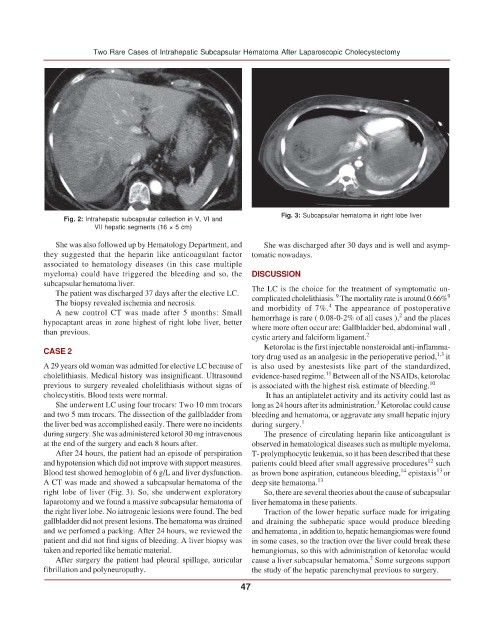
Fig. 3: Subcapsular hematoma in right lobe liver
Fig. 2: Intrahepatic subcapsular collection in V, VI and
VII hepatic segments (16 × 5 cm)
She was also followed up by Hematology Department, and She was discharged after 30 days and is well and asymp-
they suggested that the heparin like anticoagulant factor tomatic nowadays.
associated to hematology diseases (in this case multiple
myeloma) could have triggered the bleeding and so, the DISCUSSION
subcapsular hematoma liver.
The patient was discharged 37 days after the elective LC. The LC is the choice for the treatment of symptomatic un- 9
9
The biopsy revealed ischemia and necrosis. complicated cholelithiasis. The mortality rate is around 0.66%
4
A new control CT was made after 5 months: Small and morbidity of 7%. The appearance of postoperative
2
hypocaptant areas in zone highest of right lobe liver, better hemorrhage is rare ( 0.08-0-2% of all cases ), and the places
than previous. where more often occur are: Gallbladder bed, abdominal wall ,
cystic artery and falciform ligament. 2
Ketorolac is the first injectable nonsteroidal anti-inflamma-
CASE 2
1,3
tory drug used as an analgesic in the perioperative period, it
A 29 years old woman was admitted for elective LC because of is also used by anestesists like part of the standardized,
11
cholelithiasis. Medical history was insignificant. Ultrasound evidence-based regime. Between all of the NSAIDs, ketorolac
previous to surgery revealed cholelithiasis without signs of is associated with the highest risk estimate of bleeding. 10
cholecystitis. Blood tests were normal. It has an antiplatelet activity and its activity could last as
3
She underwent LC using four trocars: Two 10 mm trocars long as 24 hours after its administration. Ketorolac could cause
and two 5 mm trocars. The dissection of the gallbladder from bleeding and hematoma, or aggravate any small hepatic injury
the liver bed was accomplished easily. There were no incidents during surgery. 1
during surgery. She was administered ketorol 30 mg intravenous The presence of circulating heparin like anticoagulant is
at the end of the surgery and each 8 hours after. observed in hematological diseases such as multiple myeloma,
After 24 hours, the patient had an episode of perspiration T- prolymphocytic leukemia, so it has been described that these
12
and hypotension which did not improve with support measures. patients could bleed after small aggressive procedures such
14
13
Blood test showed hemoglobin of 6 g/L and liver dysfunction. as brown bone aspiration, cutaneous bleeding, epistaxis or
A CT was made and showed a subcapsular hematoma of the deep site hematoma. 13
right lobe of liver (Fig. 3). So, she underwent exploratory So, there are several theories about the cause of subcapsular
laparotomy and we found a massive subcapsular hematoma of liver hematoma in these patients.
the right liver lobe. No iatrogenic lesions were found. The bed Traction of the lower hepatic surface made for irrigating
gallbladder did not present lesions. The hematoma was drained and draining the subhepatic space would produce bleeding
and we perfomed a packing. After 24 hours, we reviewed the and hematoma , in addition to, hepatic hemangiomas were found
patient and did not find signs of bleeding. A liver biopsy was in some cases, so the traction over the liver could break these
taken and reported like hematic material. hemangiomas, so this with administration of ketorolac would
2
After surgery the patient had pleural spillage, auricular cause a liver subcapsular hematoma. Some surgeons support
fibrillation and polyneuropathy. the study of the hepatic parenchymal previous to surgery.
47