
Page 31 - World Journal of Laparoscopic Surgery
P. 31
Majid A Hamood
2nd group: Without laparoscopic visualization (must be seen
by surgeon, no telescope).
FIRST GROUP
The manipulation of this group is performed from inside the
abdomen under direct visualization, the maximum safety in
avoiding visceral injuries. These techniques include Maciol
needles, the Grice needle, catheter or spinal needles, the
endoclose device, and the Gor-Tex device, Reverdin, Deschamps
needles, Semm's emergency needle with adistal eyelet; the
modified Veress needle with a slitmade in the retractable brunt
tip; dental awl with aneye; prolene 2/0 on a straight needle
aided by a Veress needle; a straight needle armed with
suture;Autostitch (United states surgical), a modified Veress
needle bearing a crochet hook at the tip, veress needle loop
technique. 29
16
Grice needles Used by Stringer et al, A Grice needle (Figs
1A and B) was inserted at an angle along the side of a lateral
trocar. Under direct laparoscopic visualization, the needle was
placed through both the peritoneum and the fascia. Within the
abdomen, the suture was grasped and removed from the Grice
needle with a grasper inserted from the opposite trocar. The
Grice needle then was removed and reinserted opposite the Figs 2A to C: (A) Maciol suture needle set (B and C)
previous puncture, again at an angle along the trocar. The suture Maciol needles
was regrasped with the Grice needle and pulled out of the
abdomen. After complete removal of the trocar, the suture was
tied under direct laparoscopic visualization.
6
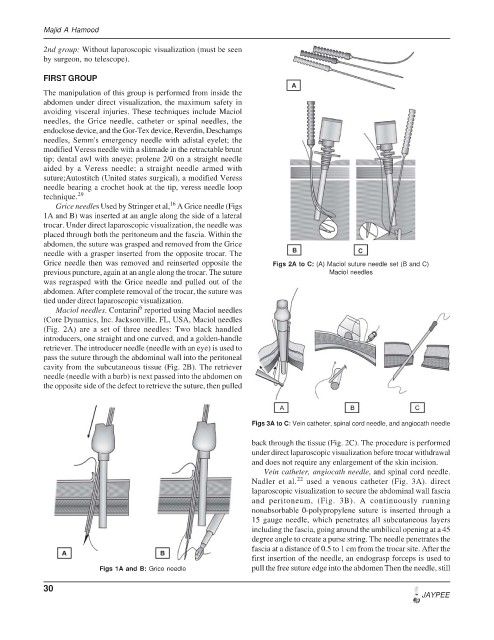
Maciol needles. Contarini reported using Maciol needles
(Core Dynamics, Inc. Jacksonville, FL, USA, Maciol needles
(Fig. 2A) are a set of three needles: Two black handled
introducers, one straight and one curved, and a golden-handle
retriever. The introducer needle (needle with an eye) is used to
pass the suture through the abdominal wall into the peritoneal
cavity from the subcutaneous tissue (Fig. 2B). The retriever
needle (needle with a barb) is next passed into the abdomen on
the opposite side of the defect to retrieve the suture, then pulled
Figs 3A to C: Vein catheter, spinal cord needle, and angiocath needle
back through the tissue (Fig. 2C). The procedure is performed
under direct laparoscopic visualization before trocar withdrawal
and does not require any enlargement of the skin incision.
Vein catheter, angiocath needle, and spinal cord needle.
22
Nadler et al. used a venous catheter (Fig. 3A). direct
laparoscopic visualization to secure the abdominal wall fascia
and peritoneum, (Fig. 3B). A continuously running
nonabsorbable 0-polypropylene suture is inserted through a
15 gauge needle, which penetrates all subcutaneous layers
including the fascia, going around the umbilical opening at a 45
degree angle to create a purse string. The needle penetrates the
fascia at a distance of 0.5 to 1 cm from the trocar site. After the
first insertion of the needle, an endograsp forceps is used to
Figs 1A and B: Grice needle pull the free suture edge into the abdomen Then the needle, still
30
JAYPEE