
Page 32 - Peer Reviewed Laparoscopic Jornal
P. 32
10.5005/jp-journals-10033-1191
Priya Bhave Chittawar et al
CASE REPORT
Hysteroscopic Findings in an Unusual Case of Adenomyosis
Priya Bhave Chittawar, Shilpa Sapre, Shilpa Bhandari
ABSTRACT fibroid uterus for myomectomy. There was no history of
Adenomyosis is the presence of ectopic endometrial glands and preceding amenorrhea.
stroma in the myometrium. It traditionally presents with pelvic Her previous cycles were prolonged with progressively
pain, menorrhagia and dysmenorrhea in the fourth or fifth decade worsening bleeding, flooding and passage of clots since
of life. Here, we present a case of adenomyosis presenting with
severe menorrhagia at the age of 23 years. 2 years. She was married for 3 years, not using any
Traditionally, adenomyosis is diagnosed in histopathologically, contraception and was desirous of conception. She was pale
in hysterectomy specimens or myometrial biopsies. Noninvasive with stable vitals. Examination revealed a uterus of 14 weeks
modalities, such as transvaginal ultrasound and magnetic in size, soft, mobile and tender with free fornices. Investi-
resonance imaging aid in diagnosis in the office before treatment
is undertaken. Office hysteroscopy is an established tool in the gations revealed Hb of 7 gm%, beta hCG <1 IU/ml, thyroid
diagnosis of abnormal uterine bleeding and infertility.While function was normal, viral screen was negative and
hysteroscopy does not have pathognomonic features of coagulation profile was normal. Transvaginal ultrasound
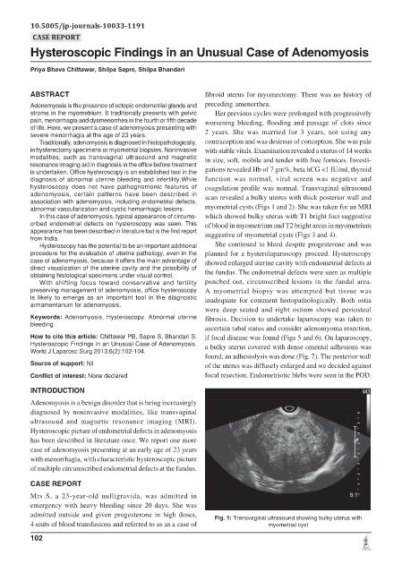
adenomyosis, certain patterns have been described in scan revealed a bulky uterus with thick posterior wall and
association with adenomyosis, including endometrial defects,
abnormal vascularization and cystic hemorrhagic lesions. myometrial cysts (Figs 1 and 2). She was taken for an MRI
In this case of adenomyosis, typical appearance of circums- which showed bulky uterus with T1 bright foci suggestive
cribed endometrial defects on hysteroscopy was seen. This of blood in myometrium and T2 bright areas in myometrium
appearance has been described in literature but is the first report suggestive of myometrial cysts (Figs 3 and 4).
from India.
Hysteroscopy has the potential to be an important additional She continued to bleed despite progesterone and was
procedure for the evaluation of uterine pathology, even in the planned for a hysterolaparoscopy proceed. Hysteroscopy
case of adenomyosis, because it offers the main advantage of showed enlarged uterine cavity with endometrial defects at
direct visualization of the uterine cavity and the possibility of
obtaining histological specimens under visual control. the fundus. The endometrial defects were seen as multiple
With shifting focus toward conservative and fertility punched out, circumscribed lesions in the fundal area.
preserving management of adenomyosis, office hysteroscopy A myometrial biopsy was attempted but tissue was
is likely to emerge as an important tool in the diagnostic inadequate for comment histopathologically. Both ostia
armamentarium for adenomyosis.
were deep seated and right ostium showed periosteal
Keywords: Adenomyosis, Hysteroscopy, Abnormal uterine fibrosis. Decision to undertake laparoscopy was taken to
bleeding.
ascertain tubal status and consider adenomyoma resection,
How to cite this article: Chittawar PB, Sapre S, Bhandari S. if focal disease was found (Figs 5 and 6). On laparoscopy,
Hysteroscopic Findings in an Unusual Case of Adenomyosis. a bulky uterus covered with dense omental adhesions was
World J Laparosc Surg 2013;6(2):102-104.
found; an adhesiolysis was done (Fig. 7). The posterior wall
Source of support: Nil of the uterus was diffusely enlarged and we decided against
Conflict of interest: None declared focal resection. Endometriotic blebs were seen in the POD.
INTRODUCTION
Adenomyosis is a benign disorder that is being increasingly
diagnosed by noninvasive modalities, like transvaginal
ultrasound and magnetic resonance imaging (MRI).
Hysteroscopic picture of endometrial defects in adenomyosis
has been described in literature once. We report one more
case of adenomyosis presenting at an early age of 23 years
with menorrhagia, with characteristic hysteroscopic picture
of multiple circumscribed endometrial defects at the fundus.
CASE REPORT
Mrs S, a 23-year-old nulligravida, was admitted in
emergency with heavy bleeding since 20 days. She was
admitted outside and given progesterone in high doses,
Fig. 1: Transvaginal ultrasound showing bulky uterus with
4 units of blood transfusions and referred to us as a case of myometrial cyst
102