
Page 7 - WJOLS - Laparoscopic Journal
P. 7
WJOLS
Laparoscopic Surgery: Results of a Modified Open Technique of Umbilical Port Insertion
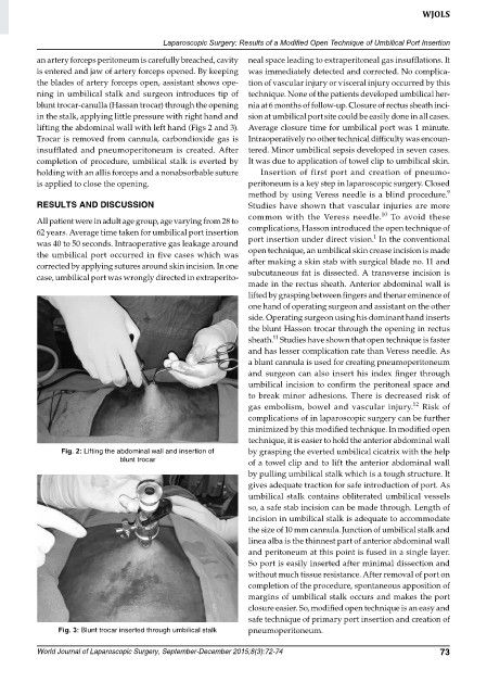
an artery forceps peritoneum is carefully breached, cavity neal space leading to extraperitoneal gas insufflations. It
is entered and jaw of artery forceps opened. By keeping was immediately detected and corrected. No complica-
the blades of artery forceps open, assistant shows ope- tion of vascular injury or visceral injury occurred by this
ning in umbilical stalk and surgeon introduces tip of technique. None of the patients developed umbilical her-
blunt trocar-canulla (Hassan trocar) through the opening nia at 6 months of follow-up. Closure of rectus sheath inci-
in the stalk, applying little pressure with right hand and sion at umbilical port site could be easily done in all cases.
lifting the abdominal wall with left hand (Figs 2 and 3). Average closure time for umbilical port was 1 minute.
Trocar is removed from cannula, carbondioxide gas is Intraoperatively no other technical difficulty was encoun-
insufflated and pneumoperitoneum is created. After tered. Minor umbilical sepsis developed in seven cases.
completion of procedure, umbilical stalk is everted by It was due to application of towel clip to umbilical skin.
holding with an allis forceps and a nonabsorbable suture Insertion of first port and creation of pneumo-
is applied to close the opening. pe ritoneum is a key step in laparoscopic surgery. Closed
9
method by using Veress needle is a blind procedure.
RESULTS AND DISCUSSION Studies have shown that vascular injuries are more
10
common with the Veress needle. To avoid these
All patient were in adult age group, age varying from 28 to
62 years. Average time taken for umbilical port insertion complications, Hasson introduced the open technique of
1
was 40 to 50 seconds. Intraoperative gas leakage around port insertion under direct vision. In the conventional
the umbilical port occurred in five cases which was open technique, an umbilical skin crease incision is made
corrected by applying sutures around skin incision. In one after making a skin stab with surgical blade no. 11 and
case, umbilical port was wrongly directed in extraperito- subcutaneous fat is dissected. A transverse incision is
made in the rectus sheath. Anterior abdominal wall is
lifted by grasping between fingers and thenar eminence of
one hand of operating surgeon and assistant on the other
side. Operating surgeon using his dominant hand inserts
the blunt Hasson trocar through the opening in rectus
11
sheath. Studies have shown that open technique is faster
and has lesser complication rate than Veress needle. As
a blunt cannula is used for creating pneumoperitoneum
and surgeon can also insert his index finger through
umbilical incision to confirm the peritoneal space and
to break minor adhesions. There is decreased risk of
12
gas embolism, bowel and vascular injury. Risk of
complications of in laparoscopic surgery can be further
minimized by this modified technique. In modified open
technique, it is easier to hold the anterior abdominal wall
Fig. 2: Lifting the abdominal wall and insertion of by grasping the everted umbilical cicatrix with the help
blunt trocar of a towel clip and to lift the anterior abdominal wall
by pulling umbilical stalk which is a tough structure. It
gives adequate traction for safe introduction of port. As
umbilical stalk contains obliterated umbilical vessels
so, a safe stab incision can be made through. Length of
incision in umbilical stalk is adequate to accommodate
the size of 10 mm cannula. Junction of umbilical stalk and
linea alba is the thinnest part of anterior abdominal wall
and peritoneum at this point is fused in a single layer.
So port is easily inserted after minimal dissection and
without much tissue resistance. After removal of port on
completion of the procedure, spontaneous apposition of
margins of umbilical stalk occurs and makes the port
closure easier. So, modified open technique is an easy and
safe technique of primary port insertion and creation of
Fig. 3: Blunt trocar inserted through umbilical stalk pneumoperitoneum.
World Journal of Laparoscopic Surgery, September-December 2015;8(3):72-74 73