
Page 46 - Journal of Laparoscopic Surgery - WALS Journal
P. 46
WJOLS
Hana Alhomoud 10.5005/jp-journals-10033-1270
CASE REPORT
Retrorectal Schwannoma
Hana Alhomoud
ABSTRACT
Schwannoma is a benign encapsulated nerve sheath tumor.
These tumors are more frequently located in the head, neck,
extremities, and trunk. Retroperitoneal pelvic localization of
schwannoma accounts for 0.5 to 5% of all cases, while the
incidence of retrorectal tumors is estimated at 1 in 40,000 to
63,000 cases in the general population, which we report here.
Keywords: Retrorectal tumors, Schwannoma, Surgery.
How to cite this article: Alhomoud H. Retrorectal Schwannoma.
World J Lap Surg 2016;9(1):44-46.
Source of support: Nil
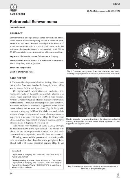
Conflict of interest: None Fig. 1: Computed tomography of the chest, abdomen, and pelvis
showing a large right lower pelvic mass, whose nature is not clear
CASE REPORT
A 50-year-old male presented with a feeling of heaviness
in the pelvic floor associated with change in bowel habits
and tenesmus for the last 5 years.
On digital rectal examination, an irreducible firm
mass posteriorly on the right side was felt. Mucosa was
intact. Rigid sigmoid scope up to 20 cm was normal.
Routine laboratory tests and tumor markers were within
normal limits. Computed tomography (CT) of the chest,
abdomen, and pelvis showed a large right lower pelvic
mass, whose nature was not clear (Fig. 1). Magnetic
resonance imaging (MRI) of the abdomen and pelvis
showed a large right presacral mass, whose appearance
suggested a neurogenic tumor (Fig. 2). Endorectal
ultrasound was done which showed a mass suggestive Fig. 2: Magnetic resonance imaging of the abdomen and pelvis
showing a large right presacral mass, whose appearance could
of sarcoma or a duplicated cyst (Fig. 3). suggest a neurogenic tumor
The patient was operated on April 2, 2012, through
an arcuate incision in the right buttock. The patient was
placed in the prone jack-knife position. An oval well-
circumscribed encapsulated mass 10 × 8 cm was removed.
Histology revealed the presence of compact spindle
cells arranged in short bundles and a peripheral lym-
phoid cuff with some germinal centers (Fig. 4). At
Consultant
Department of Surgery and Medicine, Al-Sabah Hospital
Kuwait City, Kuwait
Corresponding Author: Hana Alhomoud, Consultant
Department of Surgery and Medicine, Al-Sabah Hospital
Kuwait City, Kuwait, Phone: +96599227676, e-mail: hana_
alhomoud@hotmail.com Fig. 3: Endorectal ultrasound (showing a mass suggestive of
sarcoma or a duplicated cyst)
44