
Page 33 - wjols
P. 33
Laparoscopic Retrieval of Contraceptive Device
3,4
non-lactating women. Hypoestrogenic state with consequent
thinning of the wall of uterus and accelerated involution of the
uterus during the period of lactation could have been most likely
the causes of perforation in our first patient.
Uterine perforations are reported to mainly occur in the early
4
post-insertion period, specifically during the immediate 6 months,
but there have been case reports of perforation seen several years
5,6
after insertion. Subsequently, the IUCD can migrate into the
neighboring organs or the abdominal cavity. Trauma during the
insertion procedure itself, and along with the effect of chronic
inflammatory reaction that causes erosion of the device through
the uterine wall, can be thought to be the mechanism of IUCD
migration. Delayed symptoms are presumed to be secondary
migration with associated inflammatory process. Movements of
the omentum may be a reason of migration of the IUCD to an
adjacent organ. Migration can also be due to the growing uterus in
unintended pregnancies and tubal ectopic pregnancy. The various
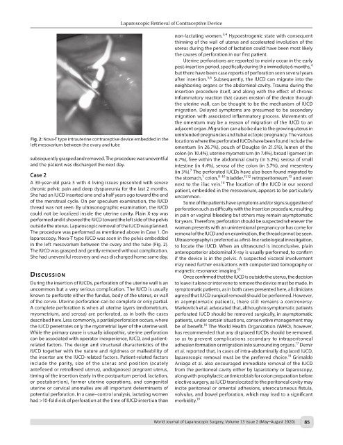
Fig. 2: Nova-T type intrauterine contraceptive device embedded in the locations where the perforated IUCDs have been found include the
left mesovarium between the ovary and tube omentum (in 26.7%), pouch of Douglas (in 21.5%), lumen of the
colon (in 10.4%), uterine myometrium (in 7.4%), broad ligament (in
subsequently grasped and removed. The procedure was uneventful 6.7%), free within the abdominal cavity (in 5.2%), serosa of small
and the patient was discharged the next day. intestine (in 4.4%), serosa of the colon (in 3.7%), and mesentery
7
(in 3%). The perforated IUCDs have also been found migrated to
Case 2 the stomach, colon, 8–10 bladder, 11,12 retroperitoneum, and even
13
1
A 39-year-old para 5 with 4 living issues presented with severe next to the iliac vein. The location of the IUCD in our second
14
chronic pelvic pain and deep dyspareunia for the last 2 months. patient, embedded in the mesovarium, appears to be particularly
She had an IUCD inserted one and a half years ago toward the end uncommon.
of the menstrual cycle. On per speculum examination, the IUCD Some of the patients have symptoms and/or signs suggestive of
thread was not seen. By ultrasonographic examination, the IUCD perforation such as difficulty with the insertion procedure, resulting
could not be localized inside the uterine cavity. Plain X-ray was in pain or vaginal bleeding but others may remain asymptomatic
performed and it showed the IUCD toward the left side of the pelvis for years. Therefore, perforation should be suspected whenever the
outside the uterus. Laparoscopic removal of the IUCD was planned. woman presents with an unintentional pregnancy or has come for
The procedure was performed as mentioned above in Case 1. On removal of the IUCD and on examination, the thread cannot be seen.
laparoscopy, Nova-T type IUCD was seen in the pelvis embedded Ultrasonography is preferred as a first-line radiological investigation,
in the left mesovarium between the ovary and the tube (Fig. 2). to locate the IUCD. When an ultrasound is inconclusive, plain
The IUCD was grasped and gently removed without complication. anteroposterior abdominal X-ray is usually performed, to confirm
She had uneventful recovery and was discharged home same day. if the device is in the pelvis. A suspected visceral involvement
may need further evaluations with computerized tomography or
dIscussIon magnetic resonance imaging. 15
Once confirmed that the IUCD is outside the uterus, the decision
During the insertion of IUCDs, perforation of the uterine wall is an to leave it alone or intervene to remove the device must be made. In
uncommon but a very serious complication. The IUCD is usually symptomatic patients, as in both cases presented here, all clinicians
known to perforate either the fundus, body of the uterus, or wall agreed that IUCD surgical removal should be performed. However,
of the cervix. Uterine perforation can be complete or only partial. in asymptomatic patients, there still remains a controversy.
A complete perforation is when all uterine layers (endometrium, Markovitch et al. advocated that, although in symptomatic patients
myometrium, and serosa) are perforated, as in both the cases perforated IUCD should be removed surgically, in asymptomatic
described here. Less commonly, a partial perforation occurs, where patients, under certain situations, conservative management may
16
the IUCD penetrates only the myometrial layer of the uterine wall. be of benefit. The World Health Organization (WHO), however,
While the primary cause is usually idiopathic, uterine perforation has recommended that any displaced IUCDs should be removed,
can be associated with operator inexperience, IUCD, and patient- so as to prevent complications secondary to intraperitoneal
17
related factors. The design and structural characteristics of the adhesion formation or migration into surrounding organs. Demir
IUCD together with the nature and rigidness or malleability of et al. reported that, in cases of intra-abdominally displaced IUCD,
18
the inserter are the IUCD-related factors. Patient-related factors laparoscopic removal must be the preferred choice. Grimaldo
include the parity, size of the uterus and position (acutely Arriaga et al. also encouraged immediate removal of the IUCD
anteflexed or retroflexed uterus), undiagnosed pregnant uterus, from the peritoneal cavity either by laparotomy or laparoscopy,
timing of the insertion (early in the postpartum period, lactation, along with prophylactic antimicrobials for colon preparation before
or postabortion), former uterine operations, and congenital elective surgery, as IUCD translocated to the peritoneal cavity may
uterine or cervical anomalies are all important determinants of incite peritoneal or omental adhesions, uterocutaneous fistula,
potential perforation. In a case–control analysis, lactating women volvulus, and bowel perforation, which may lead to a significant
had >10-fold risk of perforation at the time of IUCD insertion than morbidity. 19
World Journal of Laparoscopic Surgery, Volume 13 Issue 2 (May–August 2020) 85