
Page 26 - wjols
P. 26
Single-incision Laparoscopy-assisted Assisted Appendectomy
8
tomography was performed. Intravenous antibiotics were among the pediatric surgeons. This technique has been evolving
administered. All patients were clinically monitored for 24 hours since then and there have been several modifications in order
for resolution of clinical signs (vomiting, fever, tachycardia, right to achieve better cosmetic results, reduction in costs, shorter
iliac fossa tenderness). Patients showing clinical response within recovery period, and less hospital stay. 8,10 These newer techniques
24 hours were offered SILAA after 6 weeks. are appendectomy by laparoscopy-assisted approach, two-port
Under general anesthesia, in supine position with the patient laparoscopic approach, transumbilical single-port laparoscopic
catheterized and strapped to the operating table, an infraumbilical conventional appendectomy, and transumbilical single-incision
skin fold incision was made and deepened. Umbilical tube was laparoscopy-assisted approach. 8,10
identified and a 5 mm camera port was inserted by open Hassan’s Single-incision and single-port laparoscopic appendectomy
technique. Capnoperitoneum was created and the pressure was uses all three ports introduced through the infraumbilical incision
maintained between 8 mm Hg and 10 mm Hg. Appendix was and appendectomy is performed as in the conventional three-port
visualized. Another incision was made adjacent to the port site manner by performing endocorporeal laparoscopic appendectomy.
on the left and a 5 mm instrument was introduced through this. The single-port laparoscopic appendectomy is a recent advance
Appendicular adhesions were dissected and appendix was freed. which uses a single port with three or four internal lumens. However,
If necessary, a third incision was made to introduce an additional it requires special modified instruments—the single-incision port,
instrument to aid dissection. The tip of the appendix was held by curved instruments, and expertise; this ultimately increases the cost
6
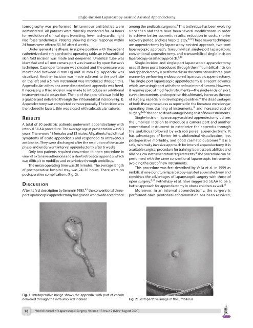
a grasper and delivered through the infraumbilical incision (Fig. 1). of surgery, especially in developing countries. The disadvantages
Appendectomy was completed extracorporeally. The incision was of both these procedures as reported in the literature were longer
11
then closed in layers. Skin was closed with subcuticular sutures. operating time, clashing of instruments, and increased cost of
surgery; 12,13 the added disadvantage being cost of new instruments.
results Single-incision laparoscopy-assisted appendectomy utilizes
the umbilical incision to introduce a camera port and another
A total of 50 pediatric patients underwent appendectomy with conventional instrument to exteriorize the appendix through
interval SILAA procedure. The average age at presentation was 9.3 the umbilicus followed by extracorporeal appendectomy. It
years. There were 18 females and 32 males. All patients had clinical has advantages of better intra-abdominal visualization, less
symptoms of acute appendicitis and responded to intravenous postoperative morbidity, and good cosmetic outcomes. It is a
8
antibiotics. They were discharged after the resolution of the acute safe, minimally invasive approach for interval appendectomy. It is
phase and underwent interval appendectomy after 6 weeks. a suitable surgical procedure for training laparoscopic abilities and
Only two patients required conversion to open procedure in also has low instrumentation requirements. The procedure can be
8
view of extensive adhesions and a short retrocecal appendix which performed with the same conventional laparoscopic instruments
was difficult to mobilize and exteriorize through umbilicus. avoiding the cost of new instruments.
The mean operating time was 30 minutes. The average length This procedure was first described by Valla et al. in 1999 as
of postoperative hospital stay was 24–36 hours. There were no umbilical one-puncture laparoscopy-assisted appendectomy and
postoperative complications (Fig. 2).
combines the advantages of laparoscopic surgery with those of
dIscussIon open surgery. 8,14 Petnehazy et al. have suggested SILAA to be a
15
better approach for appendectomy in obese children as well.
8,9
After its first description by Semm in 1983, the conventional three- Moreover, in an interval appendectomy, the surgery is
port laparoscopic appendectomy has gained worldwide acceptance performed once peritoneal contamination has been resolved,
Fig. 1: Intraoperative image shows the appendix with part of cecum
delivered through the infraumbilical incision Fig. 2: Postoperative image of the umbilicus
78 World Journal of Laparoscopic Surgery, Volume 13 Issue 2 (May–August 2020)