
Page 40 - World's Most Popular Laparoscopic Journal
P. 40
Hysteroscopic Sterilization
and posterior to the cervix. The cardinal ligament transit attempt to pass the hysteroscope may be made without the
nerve at 3 and 9 o’clock position. Uterosacral at 5 and use of tenaculum. If needed, cervical dilatation is performed.
7 o’clock position. Injection of 1% lignocaine 3 to 5 ml is Normal saline is used at body temperature and introduced
recommended at 4 and 8 or 5 and 7 to maximize the under gravity. Pressure bags may be used to maintain uterine
anesthesia while paracervical block is performed. Each step distension. Panoramic view of uterine cavity is taken, ostia
is kept informed. A nonsteroidal anti-inflammatory identified. Easier one is taken up first, which will help
suppository may help to alleviate her uterine cramps. prevent the endometrium from becoming edematous and
The OT is set with basic instruments which includes obscuring the view.
hyseroscope and diagnostic sheath, a sheath to permit The essure delivery system is passed through introducer
passage of ancillary instruments, distension media, lighting and down the working channel with tubal ostia in view.
system, and duck-billed speculum, a single-toothed The system is advanced into the proximal fallopian tube
tenaculum, dilators stand by and a paracervical kit (Fig. 4). with constant gentle forward pressure, which helps to
The procedure begins with the patient in the lithotomy prevent tubal spasm (Figs 5A to C). When the black mark
stirrups. The vulva and perineal areas are sterilely prepped is at ostia, the unit is deployed and handle of delivery device
with the iodine based solution and then sterile drapes are is stabilized against the hysteroscope. The technique involves
placed over the legs and abdomen. A pelvic examination is the thumb on essure handle, which is rotated at one click/
performed to determine the size of uterus and its orientation. sec retracting the delivery catheter and exposing the wound-
The cervix is identified. The hysteroscope is introduced down. Approximately 1 cm of insert is visible in uterine
and findings are explained to the women observing the view. cavity, i.e. small notch on wound-down insert and orange
A sterile speculum is placed on posterior vaginal wall. catheter is out and confirms proper placement. Then, button
The micro-inserts are placed using essure delivery on the handle is depressed enabling the thumb to rotate. If
system through a 5 mm hysteroscope 12 to 30 degrees, arrest of rotation and no further withdrawal of orange
which allows cannulation of fallopian tube, as it helps in catheter occurs, the procedure is complete and allows insert
forward view. With a continuous flow system, the to expand approximately 10 cm. With a counter clockwise
hysteroscope is placed under direct visualization through rotation, the delivery system is withdrawn from the catheter.
the cervix prior to dilatation. Normal saline is used during After the procedure, 3 to 8 outer coils should be visible in
placement of hysteroscope to aid visualization. An initial uterine cavity. If 18 or more, consider removal of the device.
The same procedure is repeated on contralateral side. The
mean procedure time is 9 to 13 minutes. Proper placement
of the device can be confirmed by X-ray at 11 o’clock and
1 o’clock position or ultrasound. Patency of the tube can
be ascertained by HSG after three months. Postoperatively
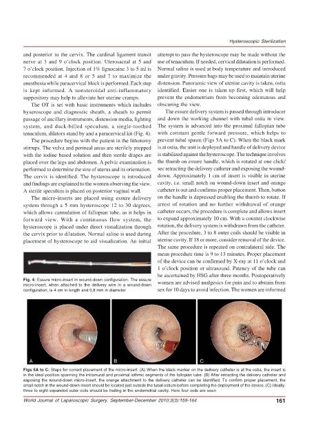
Fig. 4: Essure micro-insert in wound-down configuration. The essure women are advised analgesics for pain and to abstain from
micro-insert, when attached to the delivery wire in a wound-down
configuration, is 4 cm in length and 0.8 mm in diameter sex for 10 days to avoid infection. The women are informed
Figs 5A to C: Steps for correct placement of the micro-insert. (A) When the black marker on the delivery catheter is at the ostia, the insert is
in the ideal position spanning the intramural and proximal isthmic segments of the fallopian tube. (B) After retracting the delivery catheter and
exposing the wound-down micro-insert, the orange attachment to the delivery catheter can be identified. To confirm proper placement, the
small notch in the wound-down insert should be located just outside the tubal ostium before completing the deployment of the device. (C) Ideally,
three to eight expanded outer coils should be trailing in the endometrial cavity. Here four coils are seen
World Journal of Laparoscopic Surgery, September-December 2010;3(3):159-164 161