
Page 21 - World's Most Popular Laparoscopic Journal
P. 21
A Review of the Role of Laparoscopic Biopsy in Cases of Abdominal Lymphadenopathy
trendelenburg positions to optimally expose the site of There were no major operative and postoperative
identified lymphadenopathy. A nasogastric tube and foley complications. The average duration of hospital stay was
catheter were inserted, when appropriate both were 2.5 days ranging from 1 to 6 days.
removed at the end of surgery.
For upper abdominal procedures, a 10 mm camera DISCUSSION
port was placed slightly above the umbilicus and a Although, ultrasonography and CT are useful in identifying
5 mm working port in each midclavicular line. In abdominal lymphadenopathy, imaging findings may not
addition, a self-retaining retractor was set up to retract always be disease specific. Nodes with low density centers,
the left lobe of liver. The para-aortic nodes were biopsied although characteristic of tuberculosis, are not pathog-
by placing the camera port to the right of the midline at nomonic and nodal calcification suggestive of tuberculosis
the level of umbilicus and two working ports in the can also be observed in metastases from testicular teratoma
midline on either side. For biopsy of the external iliac and non-Hodgkin's lymphoma. Thus, the diagnosis of
11
lymph nodes, the camera port was placed at the mesenteric or retroperitoneal masses requires adequate tissue
umbilicus along with two 5 mm port in pararectus for histological evaluation as well as the possible need for
positions. After carbon dioxide insufflation begun, a immunophenotyping, cytogenetic studies, and sometimes
thorough exploration was performed. After identifying molecular genetics.
the lymph node, the peritoneum overlying the node was Image-guided biopsy is often the first line method for
carefully incised using the hook cautery. The specimen obtaining diagnostic tissue. In skilled hands ultrasono-
was grasped and isolated circumferentially from graphically-guided FNAC or CT-guided needle biopsy can
12
surrounding tissues using blunt dissection, electrocautery yield tissue samples adequate for diagnosis. While not
13
or the harmonic scalpel. The base of the node was then detailed in this review, a significant number of patients
clipped and the specimen removed. The abdomen was referred for surgical biopsy underwent successful CT-guided
irrigated and hemostasis was verified. percutaneous biopsy, avoiding the need for surgery
altogether. In a study of PFNA biopsies in 1,103 patients
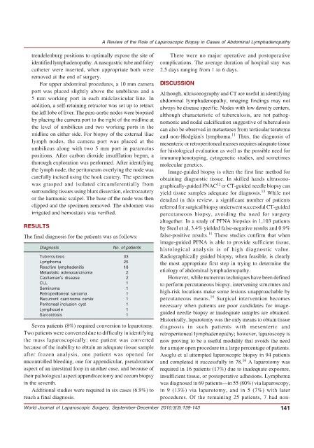
RESULTS by Steel et al, 3.4% yielded false-negative results and 0.9%
14
The final diagnosis for the patients was as follows: false-positive results. These studies confirm that when
image-guided PFNA is able to provide sufficient tissue,
Diagnosis No. of patients histological analysis is of high diagnostic value.
Tuberculosis 33 Radiographically guided biopsy, when feasible, is clearly
Lymphoma 25 the most appropriate first step in trying to determine the
Reactive lymphadenitis 18 etiology of abdominal lymphadenopathy.
Metastatic adenocarcinoma 2
Castleman's disease 2 However, while numerous techniques have been defined
CLL 1 to perform percutaneous biopsy, intervening structures and
Seminoma 1 high-risk locations make some lesions unapproachable by
Retroperitoneal sarcoma 1
15
Recurrent carcinoma cervix 1 percutaneous means. Surgical intervention becomes
Peritoneal inclusion cyst 1 necessary when patients are poor candidates for image-
Lymphocele 1 guided needle biopsy or inadequate samples are obtained.
Sarcoidosis 1
Historically, laparotomy was the only means to obtain tissue
Seven patients (8%) required conversion to laparotomy. diagnosis in such patients with mesenteric and
Two patients were converted due to difficulty in identifying retroperitoneal lymphadenopathy; however, laparoscopy is
the mass laparoscopically; one patient was converted now proving to be a useful modality that avoids the need
because of the inability to obtain an adequate tissue sample for a major open procedure in a large percentage of patients.
after frozen analysis, one patient was opened for Asoglu et al attempted laparoscopic biopsy in 94 patients
16
uncontrolled bleeding, one for appendicular, pseudotumor and completed it successfully in 78. A laparotomy was
aspect of an intestinal loop in another case, and because of required in 16 patients (17%) due to inadequate exposure,
their pathological aspect appendicectomy and cecum biopsy insufficient tissue, or postoperative adhesions. Lymphoma
in the seventh. was diagnosed in 69 patients—in 55 (80%) via laparoscopy,
Additional studies were required in six cases (6.9%) to in 9 (13%) via laparotomy, and in 5 (7%) with later
reach a final diagnosis. procedures. Of the remaining 25 patients, 7 had non-
World Journal of Laparoscopic Surgery, September-December 2010;3(3):139-143 141