
Page 7 - Journal of Laparoscopic Surgery
P. 7
WJOLS
The Camera-holding Robotic Device in Laparoscopy Surgery
to address economic concerns or lack of assistance in the used in over 175,000 procedures in over 600 hospitals around
operating room (OR); therefore, they are probably not for every the world. 7
general hospital. 3 The other device is EndoAssist (Fig. 3). EndoAssist is
During minimal access surgery, an assistant is controlling programmed to detect and follow the movements of the
the laparoscope and surgeon should be free to manipulate surgeon’s head. The surgeon wears a lightweight headband
instruments. Although the advantages of laparoscopic surgery fitted with an infrared emitter. The head position of the surgeon
are well-documented, one disadvantage is that, for optimum is detected by a receiver unit and converted into motion of the
performance, an experienced camera driver is required who can robot, so to move the view left, the surgeon simply glances to
provide the necessary views for the operating surgeon. There the left of the monitor and the camera pans round. To move the
are many drawbacks in human camera operator, especially, if view up, the surgeon looks to the top of the monitor and the
4
they are not trained. The inconvenience of laparoscopic camera follows. Movement only occurs if the surgeon is
operations lies mainly in the difficulties in mutual understanding simultaneously pressing a footswitch, thus allowing unrestricted
between the surgeon and the camera assistant who maneuvers head movements at all other times. 6
the laparoscope according to the surgeon’s instructions. Another camera-holder device was invented by Prof Mishra,
Another problem arises when the operation has to be performed India, in collaboration with Mexican engineers. ‘PMAT’, the
for many hours. In this case, the camera image tends to become name of his invention, is mechatronic assistant wih three degrees
unsteady due to fatigue of the camera assistant. The self camera- of freedom, which is made of aluminium and weighs 2.5 kg
control by the surgeon gives more stability of the laparoscopic (Fig. 4), including laparoscope and camera. This system consists
image. A robotic camera assistant, directly under surgeon’s of a harness that is placed over the surgeon’s shoulders. The
5
control, can help the surgeon control the view better. This active degree of freedom is moved in both ways using two
review is limited only in the robotic camera-holder to replace switches. To make mixed movements, the surgeon moves his/
the assistant camera-holder in laparoscopy surgery. In this her body through visual perception. This invention was helping
review, ‘Camera-holding robotic device’ term is used. Camera- the laparoscopic instrument companies to make ideal camera
holding robotic device is a robotic device that replaces the holder. 4
human assistant and ensures steady visualization of the PARAMIS (parallel robot for minimally invasive surgery)
operative field and a view which can be controlled by the was invented in Romania, which is used for laparoscope camera
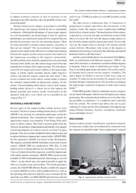
surgeon (Fig. 1). 6 positioning. The system has been built in such a way that it has
the possibility to transform it in a multiarm robot controlled
MATERIALS AND METHODS from the console. The control input allows the user to give
command in a large area for the positioning of the laparoscope
Several types of the camera-hoding robotic devices were
reviewed. The first of camera-holding robotic device is AESOP. using different interfaces: Joystick, microphone, keyboard,
8
AESOP is an acronym for automated endoscopic system for mouse and haptic device.
optimal positioning. This computerized robotic assistant for DISCUSSION
laparoscopic surgery was created by Yulun Wang, PhD, and a
team of robotic expert. They had a research grant from the Based on robotic system’s classification, such devices function
National Air and Space Administration and initially were charged as endoscopic holders that can be directed by commands from
with the development of a robotic arm for use in the US space the surgeon are classified under ‘Intern replacement’ surgical
program. This arm was later modified to hold a laparoscope and
to replace the human laparoscopic camera holder. AESOP 1000,
the first generation AESOP, was based on this development.
The surgeon controlled AESOP with either a footswitch or hand
control. AESOP 2000 was marketed in 1996 (Fig. 2) with
improvements in design and function, including voice control.
Voice activation allowed the surgeon to control the laparoscope
with simple spoken commands. AESOP 3000 system became
available in 1998. It had additional joint, functioning as a second
‘elbow’, on the robotic arm, and made it possible to apply the
robot in a broader range of procedures. The fourth generation
system, the AESOP HR (Hermes Ready), enables the surgeon
to control AESOP as well as other peripheral devices, such as
the operating table and room lights by voice command. By the
end of the year 2002, over 8000 AESOP units had been sold and Fig. 1: Laparoscopic primitive camera-holder
World Journal of Laparoscopic Surgery, September-December 2011;4(3):132-135 133