
Page 40 - Journal of Laparoscopic Surgery
P. 40
WJOLS
The Role of Laparoscopy in the Management of Mirizzi’s Syndrome: A Review of Literature
METHODS They concluded that there was a higher incidence of GBC
in patients with Mirizzi’s syndrome than in patients with
Review of literature using the SpringerLink, Google and PubMed
searches was performed and in total 148 citations were elicited. uncomplicated GSD. There were no clinical features to
Selected papers were screened for further references. Other differentiate these patients with GBC from those with Mirizzi’s
than papers in English, no other criteria for selection of literature syndrome alone, except that they were a decade older and had
was used due to the small number of articles on the syndrome. a longer duration of symptoms. In the majority, the diagnosis of
GBC was made on final histology, after cholecystectomy; hence,
FINDINGS/RESULTS this group of patients with GBC are to be treated like any other
patients with incidental GBC.
The difficult surgical management of MS is due to the presence Endoscopic retrograde cholangiopancreatography (ERCP)
of an intense fibrotic process and/or communication between is the gold standard in the diagnosis of Mirizzi’s syndrome.
the gallbladder and the common hepatic duct. Since laparoscopic
cholecystectomy became a routine procedure in the early 1990s, It delineates the cause, level, and extent of biliary obstruction,
only a few studies have been published describing their as well as ductal abnormalities, including fistulation. ERCP also
experience with the laparoscopic technique for the treatment of offers a variety of therapeutic options, such as stone extraction
MS. 9 and biliary stent placement.
M Schafer et al sampled 13,033 patients undergoing LC Percutaneous cholangiogram can provide information similar
between 1995 and 1999 and only 39 (0.3%) had MS. A total of to ERCP; however, ERCP has an additional advantage of
74% had type I MS (24/39) and five had type II MS (5/39). They identifying a low-lying cystic duct that may be missed on
concluded that MS is rarely encountered and it must be percutaneous cholangiography. Wire-guided intraductal US
recognized intraoperatively. They noted that it sometimes can provide high-resolution images of the biliary tract and
coexists with carcinoma of the gallbladder (4/39) 11% and overall adjacent structures. The diagnosis is difficult and it is more
conversion rates were 74% (24/34) for type I and 100% (5/5) for accented in third world countries where access to diagnostic
type II. techniques is limited or nonexistent. A preoperative diagnosis
Sushil K et al concluded that if not recognized is therefore made in 8 to 62.5% of all patients. 6
preoperatively, MS can result in significant morbidity and Treatment is primarily surgical. Laparoscopic surgery is the
mortality. Preoperative diagnosis may be difficult despite the standard for MS type I and II and open surgery for managing
availability of multiple imaging modalities. Ultrasonography (US), patients with types III and IV. Good short-and long-term results
CT, and magnetic resonance cholangiopancreatography with low mortality and morbidity have been reported in a number
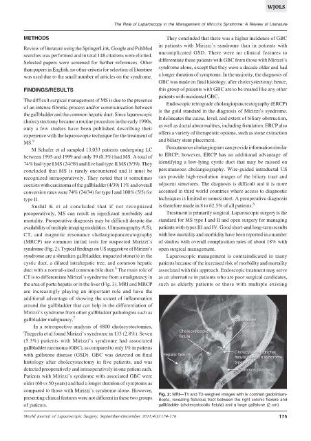
(MRCP) are common initial tests for suspected Mirizzi’s of studies with overall complication rates of about 18% with
syndrome (Fig. 2). Typical findings on US suggestive of Mirizzi’s open surgical management.
syndrome are a shrunken gallbladder, impacted stone(s) in the Laparoscopic management is contraindicated in many
cystic duct, a dilated intrahepatic tree, and common hepatic patients because of the increased risk of morbidity and mortality
5
duct with a normal-sized common bile duct. The main role of associated with this approach. Endoscopic treatment may serve
CT is to differentiate Mirizzi’s syndrome from a malignancy in as an alternative in patients who are poor surgical candidates,
the area of porta hepatis or in the liver (Fig. 3). MRI and MRCP such as elderly patients or those with multiple existing
are increasingly playing an important role and have the
additional advantage of showing the extent of inflammation
around the gallbladder that can help in the differentiation of
Mirizzi’s syndrome from other gallbladder pathologies such as
gallbladder malignancy. 7
In a retrospective analysis of 4800 cholecystectomies,
Thegeela et al found Mirizzi’s syndrome in 133 (2.8%). Seven
(5.3%) patients with Mirizzi’s syndrome had associated
gallbladder carcinoma (GBC), as compared to only 1% in patients
with gallstone disease (GSD). GBC was detected on final
histology after cholecystectomy in five patients, and was
detected preoperatively and intraoperatively in one patient each.
Patients with Mirizzi’s syndrome with associated GBC were
older (60 vs 50 years) and had a longer duration of symptoms as
compared to those with Mirizzi’s syndrome alone. However,
Fig. 2: MRI—T1 and T2-weighed images with iv contrast gadolinium-
presenting clinical features were not different in these two groups Bopta, revealing fistulous tract between the right colonic flexure and
of patients. gallbladder (cholecystocolic fistula) and a large gallstone (2 cm)
World Journal of Laparoscopic Surgery, September-December 2011;4(3):174-176 175