
Page 14 - World Journal of Laparoscopic Surgery
P. 14
Drainage of Complex Pyogenic Liver Abscess
complications, perioperative morbidity, mortality, and possible The ability to differentiate an abscess from a neoplasm at
recurrence. Twenty-two patients were managed by laparoscopic nonenhanced ultrasound is limited compared with CT or MR
drainage and 26 patients by open surgical drainage. imaging. However, if solid neoplasm starts to form necrosis, it could
All patients were subjected to full clinical assessment, laboratory be differentiated from abscess by ultrasound. 8
investigations (CBC, FBS, PP, HbA1C, creatinine, liver enzymes, By contrast enhanced CT, pyogenic liver abscess appears as
albumin and bilirubin levels, PT, PC, and INR), and at least one or two well-defined, low attenuation mass with an enhancing outer layer. It
radiological investigations (ultrasonography, computed tomography, can appear as a single nonloculated cystic collection, multiloculated
or magnetic resonance images for the abdomen and pelvis). cystic mass, solid mass, or multifocal solid lesions. 9
Abdominal ultrasonography was done in all patients and computed The characteristic imaging findings of abscess by contrast
tomography was done in 22 patients with well-defined low- enhanced CT are called (double target sign) that is seen as central
attenuation lesion that is having enhancing peripheral rim with single low attenuation cystic area surrounded by a high-density inner ring
multiloculated cystic appearance, and MRI was done in 2 patients with and a low-density outer ring. The inner layer shows early contrast
imaging feature of multiloculated cystic lesion of low T1 and high T2 enhancement with continuous enhancement at delayed phases.
signal with enhancing peripheral rim, liver abscess confirmed at right The outer layer appears of hypoattenuating with no enhancement
lobe of liver in 34 patients and at left lobe in 14 patients. Four patients in the early post contrast images then enhances in delayed phase. 6
had more than one abscess cavity. The cavity measured between 8 Another imaging findings called (cluster sign) that is seen with
cm and 23 cm in diameter. Eighteen patients had diabetes mellitus multiple small hypoattenuation abscesses aggregate and coalesce
(DM). Of the 48 patients, 9 had failed percutaneous drainage. Culture into one single large abscess cavity. Gas within lesions may be seen,
sensitivity of pus was done for all patients. either in the form of bubbles or appears as air-fluid leveling, which
Written consent form was filled by every patient after detailed is a diagnostic sign for an abscess. 10
explanation of the surgery and possible complications. At MR imaging, abscesses seen as central low T1 signal and high
T2 signal intensity, but internal signal intensity may vary depending
Patient Inclusion Criteria on the protein content. Pyogenic liver abscess appears by dynamic
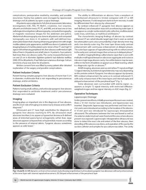
Patient having complex pyogenic liver abscess of more than 5 cm MRI contrast enhancement the same as in contrast enhanced CT,
in diameter, multilocular that is not responding to percutaneous with early enhancement of the inner layers and internal septa and
drainage, and/or antibiotics. delayed enhancement of the peripheral layer (Fig. 1).
6
Some abscesses seen surrounded with edema signal, i.e.,
Patient Exclusion Criteria appears as bright T2 signal intensity with restricted diffusion-
Patients having small, solitary, and unilocular pyogenic liver abscess weighted images and low signal intensity on ADC maps (Fig. 2). 11
that responded to antibiotic treatment and/or percutaneous
drainage were excluded. Operative Techniques
Laparoscopic Drainage
Imaging Under general anesthesia, initially pneumoperitoneum was created,
Imaging plays an important role in the diagnosis of liver abscess, then a 10 mm trochar was introduced, and laparoscope was
and the main role of imaging is to detect early disease and confirm inserted. Diagnostic laparoscopy was performed and then two 5
diagnosis. 6 mm ports were introduced according to the location of the abscess.
Ultrasound and CT have high sensitivities for diagnosis of A 10 mm port was introduced for laparoscopic intraoperative
pyogenic liver abscess reaching to 97%. By ultrasound small ultrasound. The adhesions between the liver and bowel as well as
abscesses less than 2 cm, appear as hypoechoic lesions or ill-defined the anterior abdominal wall were freed and the area where abscess
areas of distorted parenchymal echogenicity within liver, large present was exposed. Laparoscopic intraoperative ultrasound was
abscesses appears as hypoechoic or hyperechoic masses according done to detect the exact site and extent of the liver abscess, then
to the presence of internal debris. Pyogenic abscess sometimes deroofing of the abscess was done, and aspiration of the pus by the
appears as solid lesion. 7 suction catheter and samples for pus culture was taken. The cavity
Figs 1A and B: (A) MRI dynamic contrast enhancement study showing large bilobar multiloculated pyogenic liver abscesses with early enhancement
of the inner layer with internal septal enhancement; (B) Delayed enhancement of the peripheral layer
12 World Journal of Laparoscopic Surgery, Volume 13 Issue 1 (January–April 2020)