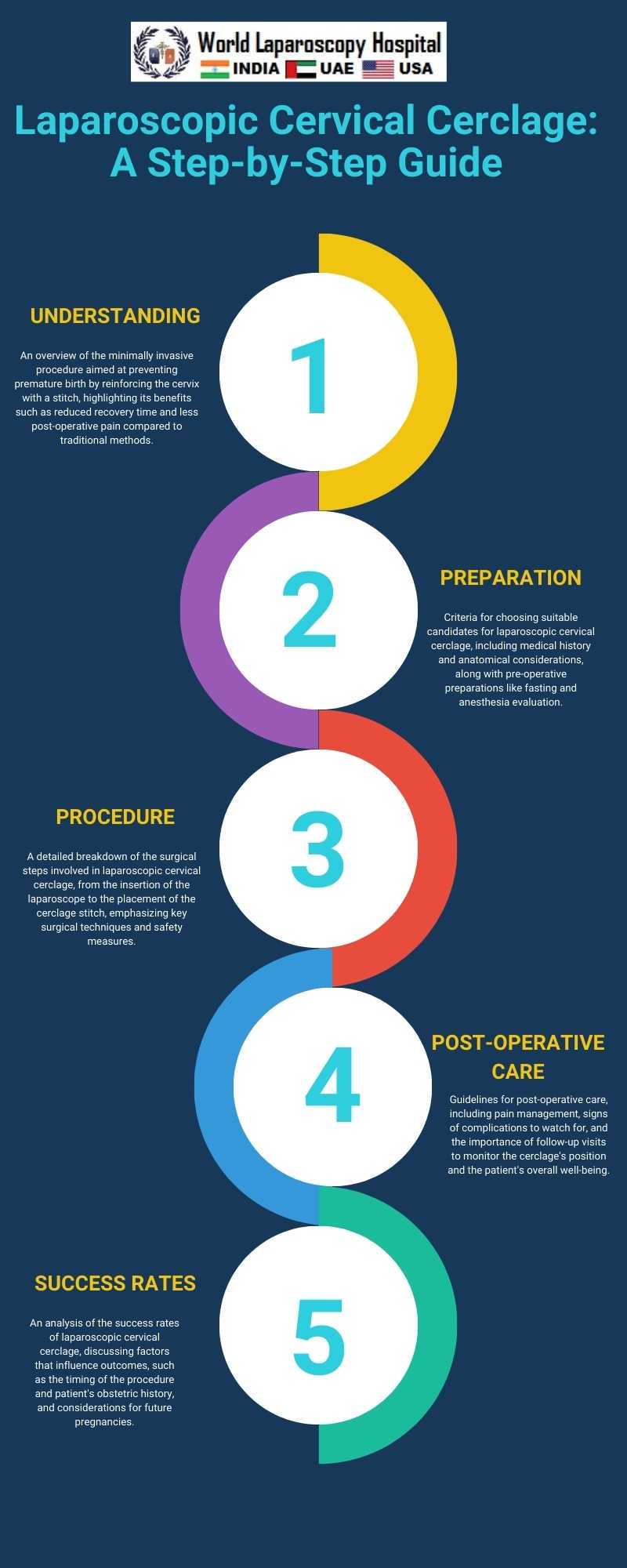
Laparoscopic Cervical Cerclage: A Step-by-Step Guide
Laparoscopic cervical cerclage is a minimally invasive surgical technique used to prevent premature birth in women with cervical insufficiency. This condition occurs when the cervix begins to dilate and efface before the pregnancy has reached term, leading to a risk of preterm birth or miscarriage. The laparoscopic approach to cervical cerclage offers several advantages over traditional methods, including reduced recovery time, decreased risk of infection, and less post-operative pain. This essay outlines a step-by-step guide to performing a laparoscopic cervical cerclage, highlighting the preoperative considerations, surgical procedure, and postoperative care.

Preoperative Considerations
Before proceeding with laparoscopic cervical cerclage, a thorough patient evaluation is essential. This includes a detailed obstetric history to identify previous preterm births or miscarriages that could suggest cervical insufficiency. Pelvic examinations and ultrasonography are crucial for assessing cervical length and funneling. Preoperative counseling should cover the risks, benefits, and potential outcomes of the procedure, ensuring the patient's informed consent.
Surgical Procedure
The laparoscopic cervical cerclage procedure is performed under general anesthesia. The patient is positioned in a dorsal lithotomy position to allow access to the uterus and cervix. The following steps outline the surgical procedure:
1. Insufflation and Trocar Placement: The abdomen is insufflated with carbon dioxide to create a working space for the laparoscopic instruments. Trocars are inserted through small incisions in the abdomen, providing access for the laparoscope and surgical tools.
2. Identifying Anatomical Landmarks: The surgeon locates key pelvic structures, including the uterus, cervix, and uterosacral ligaments. Clear visualization is essential for the accurate placement of the cerclage.
3. Dissection and Bladder Reflection: Careful dissection is carried out to separate the bladder from the anterior cervix, exposing the area where the cerclage will be placed.
4. Cerclage Placement: A non-absorbable suture is used to encircle the cervix at the level of the internal os. The suture is placed through the cervico-vaginal connective tissues, taking care to avoid the uterine vessels and the bladder.
5. Securing the Cerclage: The suture is tied securely, providing support to the cervix. The tension on the suture is adjusted to ensure it is snug but not overly tight, to prevent cervical laceration or compromised blood flow.
6. Closure: The laparoscope and instruments are removed, and the incisions are closed with sutures or surgical glue.
Postoperative Care
After the procedure, the patient is monitored for any immediate complications, such as bleeding or infection. Pain management is an important aspect of postoperative care, with most patients experiencing minimal discomfort due to the minimally invasive nature of the surgery.
Patients are advised to avoid heavy lifting and strenuous activity for a short period post-surgery. Follow-up appointments are essential for monitoring the cerclage's position and the pregnancy's progression. Ultrasound examinations are periodically performed to assess cervical length and the well-being of the fetus.
Conclusion
Laparoscopic cervical cerclage is a highly effective intervention for preventing preterm birth in women with cervical insufficiency. Its minimally invasive nature offers significant advantages over traditional cerclage methods, including shorter hospital stays, faster recovery, and reduced postoperative discomfort. By following a meticulous step-by-step approach, surgeons can ensure optimal outcomes for their patients, contributing to the successful management of high-risk pregnancies.
2 COMMENTS
Dr. Ankita Jain
#1
Feb 14th, 2024 8:14 am
Laparoscopic cervical cerclage effectively prevents preterm birth in cervical insufficiency. Its minimally invasive approach offers benefits like shorter hospital stays and faster recovery, enhancing patient comfort and contributing to successful management of high-risk pregnancies.
Dr. Baljeet Kaur
#2
Feb 27th, 2024 5:32 pm
Laparoscopic cervical cerclage is a superior intervention for preventing preterm birth in cervical insufficiency. Its minimally invasive nature yields benefits like shorter hospital stays, faster recovery, and less discomfort, ensuring optimal outcomes for high-risk pregnancies.
| Older Post | Home | Newer Post |


