Advances in Surgical Techniques: Laparoscopic Heller Myotomy for Esophageal Achalasia
Advances in Surgical Techniques: Laparoscopic Heller Myotomy for Esophageal Achalasia
Introduction:
Esophageal achalasia is a rare but debilitating disorder that affects the esophagus, making it difficult for patients to swallow food and liquids. This condition is characterized by the failure of the lower esophageal sphincter (LES) to relax properly, leading to impaired food passage into the stomach. Over the years, several surgical techniques have been developed to treat esophageal achalasia, with laparoscopic Heller myotomy emerging as a significant advancement in this field. In this article, we will explore the evolution of surgical techniques for esophageal achalasia, with a focus on the groundbreaking developments associated with laparoscopic Heller myotomy.
Understanding Esophageal Achalasia
Before delving into the surgical advancements, it's crucial to understand the pathophysiology and clinical presentation of esophageal achalasia. This condition often presents with symptoms such as dysphagia (difficulty swallowing), regurgitation, chest pain, and weight loss. These symptoms can significantly impact a patient's quality of life and nutritional status.
The primary underlying problem in esophageal achalasia is the dysfunction of the LES, a muscular ring that separates the esophagus from the stomach. In healthy individuals, the LES relaxes to allow the passage of food into the stomach. However, in achalasia patients, the LES remains contracted, creating a functional obstruction. This leads to the accumulation of food and liquids in the esophagus, causing the characteristic symptoms.
Historical Perspective on Surgical Treatment
The surgical treatment of esophageal achalasia has a long history, with various approaches developed over the years. One of the earliest surgical techniques involved the division of the LES through an open surgical procedure, known as the Heller myotomy. While this procedure was effective in relieving symptoms, it was associated with a significant recovery period and postoperative pain.
The Advent of Laparoscopic Heller Myotomy
The development of laparoscopic surgery in the late 20th century revolutionized many surgical fields, including the treatment of esophageal achalasia. Laparoscopic Heller myotomy, also known as laparoscopic esophagomyotomy, was introduced as a minimally invasive alternative to the traditional open approach. This procedure involves making small incisions in the abdomen and using specialized instruments to dissect and divide the LES muscle, allowing for its relaxation.
One of the key advantages of laparoscopic Heller myotomy is its minimally invasive nature. Compared to open surgery, patients experience less postoperative pain, shorter hospital stays, and faster recovery times. Additionally, the smaller incisions result in improved cosmetic outcomes. This procedure has become the gold standard for the surgical treatment of esophageal achalasia.
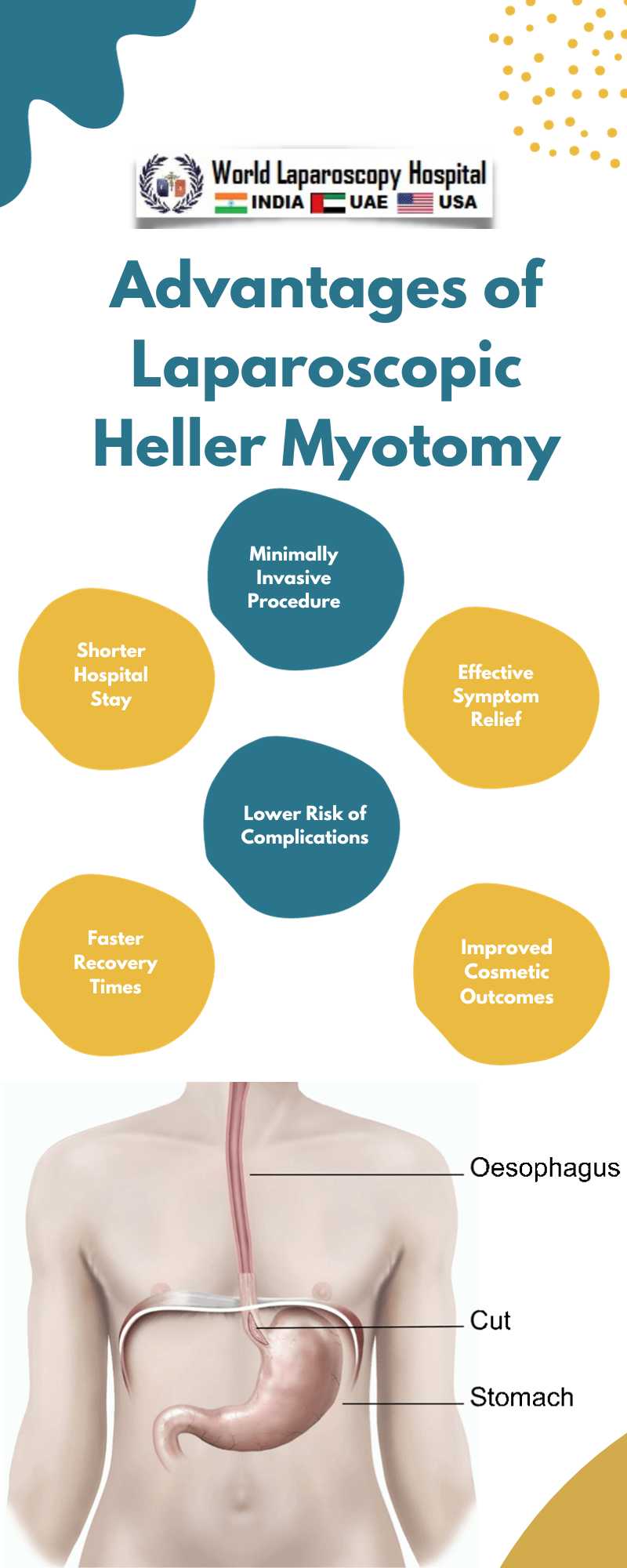
Advantages of Laparoscopic Heller Myotomy
1. Minimally Invasive: Laparoscopic Heller myotomy is performed through small incisions, reducing trauma to the abdominal wall and minimizing postoperative pain.
2. Shorter Hospital Stay: Patients undergoing laparoscopic Heller myotomy typically have shorter hospital stays compared to open surgery.
3. Faster Recovery: The minimally invasive approach allows for quicker recovery, enabling patients to return to their normal activities sooner.
4. Improved Cosmesis: The smaller incisions result in better cosmetic outcomes, with less scarring.
5. Efficacy: Laparoscopic Heller myotomy has been shown to effectively relieve symptoms in the majority of esophageal achalasia patients.
6. Lower Complication Rates: Studies have demonstrated lower complication rates associated with laparoscopic Heller myotomy compared to open surgery.
Techniques and Variations
As laparoscopic Heller myotomy gained popularity, surgeons began to refine the technique and explore variations to optimize outcomes. One such variation is the addition of an antireflux procedure, such as a partial fundoplication, to prevent gastroesophageal reflux disease (GERD) in some patients. This modification has shown promise in reducing the risk of postoperative reflux while maintaining the benefits of symptom relief.
Additionally, advancements in surgical instruments and technology have further improved the precision and safety of laparoscopic Heller myotomy. High-definition cameras, robotic-assisted surgery, and advanced energy devices have all contributed to enhanced surgical outcomes.
Patient Selection and Preoperative Evaluation
Successful outcomes in laparoscopic Heller myotomy depend on careful patient selection and thorough preoperative evaluation. Patients must undergo a comprehensive assessment to confirm the diagnosis of esophageal achalasia and evaluate their overall health.
Diagnostic Modalities
1. Esophageal Manometry: This test measures the pressure in the esophagus and LES, confirming the absence of normal relaxation in achalasia.
2. Barium Swallow: A barium contrast study helps visualize the esophagus and assess the extent of dilation and retention of contrast material.
3. Endoscopy: Upper endoscopy can rule out other esophageal conditions and may reveal food or fluid retention in the esophagus.
Patient Evaluation
In addition to confirming the diagnosis, preoperative evaluation includes assessing the patient's general health, comorbidities, and the presence of GERD. Patients with achalasia often have dilated esophagi, which may require additional interventions such as pneumatic dilation or botulinum toxin injection before surgery.
Surgical Technique
Laparoscopic Heller myotomy is typically performed under general anesthesia. The surgeon makes several small incisions in the upper abdomen, through which trocars (thin tubes) are inserted to provide access for specialized instruments and a laparoscope. The key steps of the procedure include:
1. Exposure and Dissection: The surgeon identifies the LES and carefully dissects the muscle layers to expose the inner circular muscle layer of the esophagus.
2. Myotomy: Using electrocautery or other energy devices, the surgeon performs a myotomy by dividing the circular muscle fibers of the esophagus and extending it onto the stomach for several centimeters. The goal is to create a controlled, partial thickness division of the muscle layer, allowing for relaxation of the LES.
3. Dor Fundoplication (Optional): In some cases, a partial fundoplication may be performed to prevent postoperative reflux. This involves wrapping a portion of the stomach around the lower esophagus.
4. Closure and Recovery: The trocars are removed, and the small incisions are closed. Patients are typically observed in the hospital for a short period before being discharged.
Postoperative Care and Follow-Up
After laparoscopic Heller myotomy, patients are advised to follow a specific diet to allow the surgical site to heal gradually. This typically involves consuming soft or liquid foods for a few weeks before transitioning to a regular diet. Patients are also instructed to avoid heavy lifting and strenuous activities during the initial recovery period.
Follow-up appointments are crucial to monitor progress and address any concerns. Patients may undergo postoperative tests, such as barium swallows and esophageal manometry, to assess the effectiveness of the procedure. Long-term follow-up is essential to ensure that symptom relief is sustained and to address any potential complications.
Outcomes and Complications
The success rates of laparoscopic Heller myotomy are generally high, with most patients experiencing significant symptom relief. Dysphagia and regurgitation are typically improved or resolved in the majority of cases. However, like any surgical procedure, laparoscopic Heller myotomy is not without potential complications, which may include:
1. GERD: While laparoscopic Heller myotomy can improve reflux symptoms in some patients, it can also lead to an increased risk of GERD in others. This is why the addition of an antireflux procedure is considered in certain cases.
2. Perforation: During the myotomy, there is a risk of unintended perforation of the esophagus or stomach. This is typically managed intraoperatively, but it may require additional interventions.
3. Infection: As with any surgery, there is a risk of infection at the incision sites or within the abdominal cavity.
4. Bleeding: In rare cases, bleeding may occur during or after surgery and may require intervention.
5. Gas Bloat Syndrome: Some patients may experience excessive gas buildup in the stomach, leading to discomfort and bloating.
6. Persistent Dysphagia: While laparoscopic Heller myotomy is effective for most patients, some may continue to experience mild dysphagia postoperatively.
Conclusion:
Laparoscopic Heller myotomy has emerged as a groundbreaking advancement in the surgical treatment of esophageal achalasia. Its minimally invasive nature, shorter recovery times, and improved patient outcomes have made it the preferred choice for many patients and surgeons. With ongoing research and technological innovations, the procedure continues to evolve, offering hope for even better outcomes and fewer complications in the future.
As surgical techniques continue to advance, it is crucial for healthcare providers to stay updated on the latest developments to provide the best possible care to patients with esophageal achalasia. Additionally, ongoing collaboration between surgeons, gastroenterologists, and other specialists is essential to ensure a multidisciplinary approach to the management of this challenging condition.
In conclusion, laparoscopic Heller myotomy represents a significant milestone in the treatment of esophageal achalasia, offering hope and improved quality of life for patients who once faced the challenges of this debilitating disorder.
Introduction:
Esophageal achalasia is a rare but debilitating disorder that affects the esophagus, making it difficult for patients to swallow food and liquids. This condition is characterized by the failure of the lower esophageal sphincter (LES) to relax properly, leading to impaired food passage into the stomach. Over the years, several surgical techniques have been developed to treat esophageal achalasia, with laparoscopic Heller myotomy emerging as a significant advancement in this field. In this article, we will explore the evolution of surgical techniques for esophageal achalasia, with a focus on the groundbreaking developments associated with laparoscopic Heller myotomy.
Understanding Esophageal Achalasia
Before delving into the surgical advancements, it's crucial to understand the pathophysiology and clinical presentation of esophageal achalasia. This condition often presents with symptoms such as dysphagia (difficulty swallowing), regurgitation, chest pain, and weight loss. These symptoms can significantly impact a patient's quality of life and nutritional status.
The primary underlying problem in esophageal achalasia is the dysfunction of the LES, a muscular ring that separates the esophagus from the stomach. In healthy individuals, the LES relaxes to allow the passage of food into the stomach. However, in achalasia patients, the LES remains contracted, creating a functional obstruction. This leads to the accumulation of food and liquids in the esophagus, causing the characteristic symptoms.
Historical Perspective on Surgical Treatment
The surgical treatment of esophageal achalasia has a long history, with various approaches developed over the years. One of the earliest surgical techniques involved the division of the LES through an open surgical procedure, known as the Heller myotomy. While this procedure was effective in relieving symptoms, it was associated with a significant recovery period and postoperative pain.
The Advent of Laparoscopic Heller Myotomy
The development of laparoscopic surgery in the late 20th century revolutionized many surgical fields, including the treatment of esophageal achalasia. Laparoscopic Heller myotomy, also known as laparoscopic esophagomyotomy, was introduced as a minimally invasive alternative to the traditional open approach. This procedure involves making small incisions in the abdomen and using specialized instruments to dissect and divide the LES muscle, allowing for its relaxation.
One of the key advantages of laparoscopic Heller myotomy is its minimally invasive nature. Compared to open surgery, patients experience less postoperative pain, shorter hospital stays, and faster recovery times. Additionally, the smaller incisions result in improved cosmetic outcomes. This procedure has become the gold standard for the surgical treatment of esophageal achalasia.
Advantages of Laparoscopic Heller Myotomy
1. Minimally Invasive: Laparoscopic Heller myotomy is performed through small incisions, reducing trauma to the abdominal wall and minimizing postoperative pain.
2. Shorter Hospital Stay: Patients undergoing laparoscopic Heller myotomy typically have shorter hospital stays compared to open surgery.
3. Faster Recovery: The minimally invasive approach allows for quicker recovery, enabling patients to return to their normal activities sooner.
4. Improved Cosmesis: The smaller incisions result in better cosmetic outcomes, with less scarring.
5. Efficacy: Laparoscopic Heller myotomy has been shown to effectively relieve symptoms in the majority of esophageal achalasia patients.
6. Lower Complication Rates: Studies have demonstrated lower complication rates associated with laparoscopic Heller myotomy compared to open surgery.
Techniques and Variations
As laparoscopic Heller myotomy gained popularity, surgeons began to refine the technique and explore variations to optimize outcomes. One such variation is the addition of an antireflux procedure, such as a partial fundoplication, to prevent gastroesophageal reflux disease (GERD) in some patients. This modification has shown promise in reducing the risk of postoperative reflux while maintaining the benefits of symptom relief.
Additionally, advancements in surgical instruments and technology have further improved the precision and safety of laparoscopic Heller myotomy. High-definition cameras, robotic-assisted surgery, and advanced energy devices have all contributed to enhanced surgical outcomes.
Patient Selection and Preoperative Evaluation
Successful outcomes in laparoscopic Heller myotomy depend on careful patient selection and thorough preoperative evaluation. Patients must undergo a comprehensive assessment to confirm the diagnosis of esophageal achalasia and evaluate their overall health.
Diagnostic Modalities
1. Esophageal Manometry: This test measures the pressure in the esophagus and LES, confirming the absence of normal relaxation in achalasia.
2. Barium Swallow: A barium contrast study helps visualize the esophagus and assess the extent of dilation and retention of contrast material.
3. Endoscopy: Upper endoscopy can rule out other esophageal conditions and may reveal food or fluid retention in the esophagus.
Patient Evaluation
In addition to confirming the diagnosis, preoperative evaluation includes assessing the patient's general health, comorbidities, and the presence of GERD. Patients with achalasia often have dilated esophagi, which may require additional interventions such as pneumatic dilation or botulinum toxin injection before surgery.
Surgical Technique
Laparoscopic Heller myotomy is typically performed under general anesthesia. The surgeon makes several small incisions in the upper abdomen, through which trocars (thin tubes) are inserted to provide access for specialized instruments and a laparoscope. The key steps of the procedure include:
1. Exposure and Dissection: The surgeon identifies the LES and carefully dissects the muscle layers to expose the inner circular muscle layer of the esophagus.
2. Myotomy: Using electrocautery or other energy devices, the surgeon performs a myotomy by dividing the circular muscle fibers of the esophagus and extending it onto the stomach for several centimeters. The goal is to create a controlled, partial thickness division of the muscle layer, allowing for relaxation of the LES.
3. Dor Fundoplication (Optional): In some cases, a partial fundoplication may be performed to prevent postoperative reflux. This involves wrapping a portion of the stomach around the lower esophagus.
4. Closure and Recovery: The trocars are removed, and the small incisions are closed. Patients are typically observed in the hospital for a short period before being discharged.
Postoperative Care and Follow-Up
After laparoscopic Heller myotomy, patients are advised to follow a specific diet to allow the surgical site to heal gradually. This typically involves consuming soft or liquid foods for a few weeks before transitioning to a regular diet. Patients are also instructed to avoid heavy lifting and strenuous activities during the initial recovery period.
Follow-up appointments are crucial to monitor progress and address any concerns. Patients may undergo postoperative tests, such as barium swallows and esophageal manometry, to assess the effectiveness of the procedure. Long-term follow-up is essential to ensure that symptom relief is sustained and to address any potential complications.
Outcomes and Complications
The success rates of laparoscopic Heller myotomy are generally high, with most patients experiencing significant symptom relief. Dysphagia and regurgitation are typically improved or resolved in the majority of cases. However, like any surgical procedure, laparoscopic Heller myotomy is not without potential complications, which may include:
1. GERD: While laparoscopic Heller myotomy can improve reflux symptoms in some patients, it can also lead to an increased risk of GERD in others. This is why the addition of an antireflux procedure is considered in certain cases.
2. Perforation: During the myotomy, there is a risk of unintended perforation of the esophagus or stomach. This is typically managed intraoperatively, but it may require additional interventions.
3. Infection: As with any surgery, there is a risk of infection at the incision sites or within the abdominal cavity.
4. Bleeding: In rare cases, bleeding may occur during or after surgery and may require intervention.
5. Gas Bloat Syndrome: Some patients may experience excessive gas buildup in the stomach, leading to discomfort and bloating.
6. Persistent Dysphagia: While laparoscopic Heller myotomy is effective for most patients, some may continue to experience mild dysphagia postoperatively.
Conclusion:
Laparoscopic Heller myotomy has emerged as a groundbreaking advancement in the surgical treatment of esophageal achalasia. Its minimally invasive nature, shorter recovery times, and improved patient outcomes have made it the preferred choice for many patients and surgeons. With ongoing research and technological innovations, the procedure continues to evolve, offering hope for even better outcomes and fewer complications in the future.
As surgical techniques continue to advance, it is crucial for healthcare providers to stay updated on the latest developments to provide the best possible care to patients with esophageal achalasia. Additionally, ongoing collaboration between surgeons, gastroenterologists, and other specialists is essential to ensure a multidisciplinary approach to the management of this challenging condition.
In conclusion, laparoscopic Heller myotomy represents a significant milestone in the treatment of esophageal achalasia, offering hope and improved quality of life for patients who once faced the challenges of this debilitating disorder.
1 COMMENTS
Dr. Diwakar
#1
Nov 26th, 2023 9:08 am
Esophageal achalasia, a rare yet debilitating disorder affecting swallowing, finds relief in laparoscopic Heller myotomy. This article explores the evolution of surgical techniques, emphasizing the groundbreaking developments associated with this significant advancement.
| Older Post | Home | Newer Post |
