
Page 8 - Journal of Laparoscopic Surgery
P. 8
Nidhi Jain et al.
was done. In women with obesity and previous surgeries, site. Two ancillary 5 mm trocars were placed as shown in
laparoscopy was preferred. Few cases with uterine size Figure 1A. In cases with the large uterus or big fibroid,
> 12 weeks and fibroid size of 8 to 10 cm have also been another 5mm trocar was placed on the lateral side for
operated laparoscopically. uterine manipulation by introducing myoma screw.
The data was collected in form of a demographic The round ligament was first cauterized with bipolar
profile, an indication of surgery, complications observed forceps and then cut with Enseal forceps. Similarly, fallopian
during surgery and postoperative complications. The tube and ovarian ligament were also cauterized and cut.
comparison was done between two groups, women After cutting the fundal structures, the vesicouterine
undergoing TAH (group I) and women undergoing TLH fold of peritoneum was opened by the harmonic blade in
(group II). A p value < 0.05 was considered significant. the central part of the lower uterine segment. After that,
bladder dissection is continued in either direction and
Surgical Technique bladder is pushed downwards. During this step, a cup
After informed consent, the patient was taken for surgery. of the uterine manipulator is pushed inside to locate the
Surgery was done under general anesthesia with end right cleavage plane (Fig. 1B).
tracheal intubation. The patient was placed in the dorsal After careful skeletonization, the uterine artery was
lithotomy position. After per vaginum examination, the coagulated with bipolar forceps and cut with scissors or
uterine manipulator (Marva’s) was placed and Foley harmonic blade (Fig. 1C). The uterosacral ligaments were
urinary catheter was inserted. then coagulated and sectioned, by harmonic.
After creating CO pneumoperitoneum with Veress Lastly, circular colpotomy was then done by using
2
needle, a 10 mm trocar was placed at the supra-umbilical the unipolar hook (Fig. 1D) and the uterus was removed
A B
C D
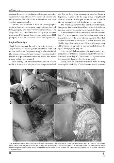
Figs 1A to D: Captional 10mm port and two ancillary 5 mm ports; (B) Laparo-
Figs 1A and D: (A) Laparoscopic image showing placem e nt o f s u p r a - u m b i l i c
scopic image showing bladder dissection via opening of vesico-uterine fold of peritoneum by harmonic blade; (C) Laparoscopic image
showing coagulation of uterine artery followed by cutting by harmonic forceps; (D) Laparoscopic image showing circular colpotomy
by unipolar hook
60