
Page 31 - WJOLS - World Journal of Laparoscopic Surgery
P. 31
Laparoscopic Conservative Treatment and Laparoscopic Salpingotomy
Table 1: Risk factors for ectopic pregnancy
Risk factor Odds ratio
High risk
Previous ectopic pregnancy 9.3–47
Previous tubal surgery 6.0–11.5
Tubal ligation 3.0–139
Tubal pathology 3.5–25
In utero DES exposure 2.4–13
Current IUD use 1.1–45
Moderate risk
Infertility 1.1–28
Previous cervicitis (gonorrhea, chlamydia) 2.8–3.7
History of pelvic inflammatory disease 2.1–3.0
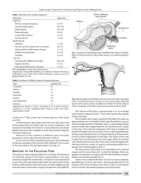
Multiple sexual partners 1.4–4.8 Fig. 1: Anatomy of the fallopian tube (modified from Netter FH. Netter
Smoking 2.3–3.9 Atlas of Human Anatomy. 3rd ed. New Jersey: Icon Learning Systems;
Low risk 2003)
Previous pelvic/abdominal surgery 0.93–3.8
Vaginal douching 1.1–3.1
Early age of intercourse (<18 years) 1.1–2.5
DES, diethylstilbestrol; IUD, intrauterine device
Adapted from: Ankum WM, Mol BWJ, et al. Fertil Steril 1996;65:1093; Murray
H, Baakdah H, et al. CMAJ 2005;173:905 and Bouyer J, Coste J, et al. Am J
Epidemiol 2003;157:185
Table 2: Incidence of different types of ectopic pregnancy
Type Incidence (%)
Ampullary 70
Isthmic 12
Fimbrial 1.1
Interstitial 2.4
Ovarian 3.2 Fig. 2: Blood supply to the fallopian tube. A cascade of vessels originating
Intra-abdominal 1.3 from an arcuate formed by a branch of the ovarian artery and tubal
Cervical <1 branch of the uterine artery (modified from Netter FH. Netter Atlas of
Adapted from: Bouyer J, Coste J, Fernandez H, et al. Sites of ectopic Human Anatomy. 3rd ed. New Jersey: Icon Learning Systems; 2003)
pregnancy: a 10 year population-based study of 1,800 cases. Hum
Reprod 2002;17:3224 The isthmus of the tube is approximately 4–6 cm in length
and its lumen is approximately 1–2 mm until it gets to the ampulla
10
10,000 IU/L). The overall rate of tubal rupture in this series where it enlarges.
was 18%. The ampulla is the longest segment of the tube and makes up
Prompt diagnosis and proper treatment may also play a role approximately two-thirds of the total length. Beneath the mucosa
in the preservation of fertility after an ectopic pregnancy. The of the ampullary portion of the tube, there is a series of large blood
increased knowledge of risk factors among clinicians and proper vessels mostly veins originating from the uterine/ovarian supply
patient education have enabled an early and accurate diagnosis to the tube. These become engorged at the time of ovulation to
of ectopic pregnancy. bring the fimbriae closer to the ovary. They can also be problematic
Awareness of the incidence of different types of ectopic during surgical treatment for an ectopic pregnancy. These vessels
pregnancy is most critical for early detection (Table 2). travel in a thick longitudinal muscle layer. The lumen of the tube
In one series of 1,800 surgically treated cases, the distribution is wider here and the mucosa has more rugae, which are covered
of sites was ampullary (70%), isthmic (12%), fimbrial (11.1%), ovarian with ciliated and secretory cells. These cells may be damaged with
3
(3.2%), interstitial (2.4%), and abdominal (1.3%) (Fig. 1). infection, previous ectopic or surgery predisposing patients to a
greater risk of tubal pregnancy (Fig. 1).
The final portion of the tube is the infundibulum; it is funnel
AnAtomy of the fAllopIAn tube shaped and its most distal end is called the fimbriae. There are
The oviduct or tube is approximately 10–12 cm long. The intramural greater concentrations of ciliary cells here that facilitate transport
or interstitial portion of the tube is approximately 1 cm long, of the ovum into the ampulla (Fig. 2).
traverses through the myometrium, and opens in the endometrial Studies that combined the level of serum β-human chorionic
cavity. This is the opening through which the sperm travel to the gonadotropin (β-hCG) and pelvic ultrasonography led to the
oviduct and the embryo enters the cavity. It is also a highly vascular concept of the discriminatory zone (level of serum β-hCG above
area and makes conservative surgical management more difficult. which a normal intrauterine pregnancy should be seen).
World Journal of Laparoscopic Surgery, Volume 11 Issue 3 (September–December 2018) 139