
Page 93 - World Journal of Laparoscopic Surgery
P. 93
Laparoscopic Management of Uncommon Presentations of Ectopic Pregnancy
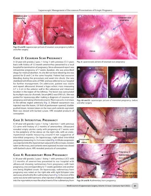
Figs 2A and B: Laparoscopic picture of cesarean scar pregnancy before
and after surgery
cAse 2: cesAreAn scAr PregnAncy
A 32-year-old gravida 2 para 1 living 1 with previous CS 3 years Fig. 3: Laparoscopic picture of cesarean scar pregnancy
back with history of 1.5 month amenorrhea presented to a local
hospital for termination of pregnancy. Since ultrasound report was
intrauterine pregnancy of 7 weeks duration, she was prescribed
drugs for medical abortion. As she did not have bleeding she was
posted for D and C in the same hospital. Patient had excessive
bleeding during the procedure and went into shock. She was
stabilized with three units of PRBC and was referred to our hospital
for further management. On admission, patient was stable
and repeat ultrasound showed a hypo echoic mass measuring
4.7 × 4 cm in the anterior wall in the subserosal and intramural
location in the region of the isthmus. The lesion was surrounded
by multiple vascular channels. Serum βHCG was 6700 U/L. She was
posted for laparoscopy after making a diagnosis of cesarean scar
pregnancy and taking informed consent. There was a 4 × 2 cm mass Figs 4A and B: Laparoscopic picture of interstitial pregnancy: before
in the isthmic region anteriorly (Fig. 3). Diluted vasopressin was and after surgery
injected near the lesion, UV fold of peritoneum opened, bladder
pushed down, incision taken on the mass and contents aspirated.
Rent was closed with barbed suture. HPE revealed products of
conception.
cAse 3: InterstItIAl PregnAncy
A 30-year-old gravida 3 para 1 living 1 abortion 1 with previous
CS came with history of 2 months of amenorrhea. Ultrasound
revealed empty uterine cavity with pregnancy of 7 weeks seen
to the periphery of the uterus on the right side, with an endo-
myometrial mantle measuring around 4 mm suggestive of
interstitial pregnancy. On laparoscopy, right-sided interstitial
pregnancy measuring 4 × 5 cm was noted (Fig. 4). Dilute vasopressin
was injected into the myometrium adjacent to the ectopic, incision
taken on the mass, and contents were aspirated. Incision was closed
with barbed suture. HPE revealed products of conception.
cAse 4: rudImentAry Horn PregnAncy
A 36-year-old gravida 2 para 1 living 1 with previous LSCS with
2.5 months of amenorrhea presented to our hospital with
ultrasound showing rudimentary horn pregnancy with twin
pregnancy, one corresponding to 11 weeks gestation and another
one being blighted ovum. On laparoscopy, rudimentary horn
pregnancy was noted on the right side with right fallopian tube
and ovary attached to the rudimentary horn (Fig. 5). Excision of the
same was done with harmonic after injection of dilute vasopressin
into the myometrium near the attachment of the rudimentary horn Figs 5A and B: Rudimentary horn pregnancy
World Journal of Laparoscopic Surgery, Volume 15 Issue 1 (January–April 2022) 91