
Page 49 - World Journal of Laparoscopic Surgery
P. 49
Postoperative Acute Pancreatitis in a Patient Who Underwent Laparoscopic Cholecystectomy
Table 1: Blood workup of the patient during initial visits to ED
Labs Hg WBC Total bilirubin AST ALP
3
During initial visit to ED—preoperative Cholecystectomy 11.8 g/dL 6.2 × 10 /mm 3 2.70 mg/dL 1000 units/L 177 IU/L
3
During postoperative day #12 visit to ED—acute pancreatitis bout. 13.6 g/dL 17.7 × 10 /mm 3 2.60 mg/dL 442 units/L 740 IU/L
1,2
performed in those over 40 years of age. However, 90% of patients
with cholelithiasis between 18 and 49 years are operated on by
8
LC. There are many possible etiologies for AP such as alcoholism,
medications, cystic fibrosis, hypercalcemia, hypertriglyceridemia,
1,2
and trauma. After ruling out these causes the patient recently
operated for cholecystitis stands the next risk factor.
The patient was operated satisfactorily without any evidence
of complication. It was seen that the patient presented a case of
acute cholecystitis, which is considered a risk factor for conversion
9
to open surgery; however, our patient did not present this common
complication. A study found that mild thickened (from 2–4 mm)
gallbladder had more risk to present complications compared with
10
normal wall thickness, 53.1 vs 10.5%. In the case of our patient, she
has a 2.5 mm thickened gallbladder. Regarding the stones found in
the gallbladder, the stones were small and multiple. Some studies
mention that the presence of smaller stones predisposes a greater
11
risk of later pancreaticobiliary events. To rule out bile leakage in
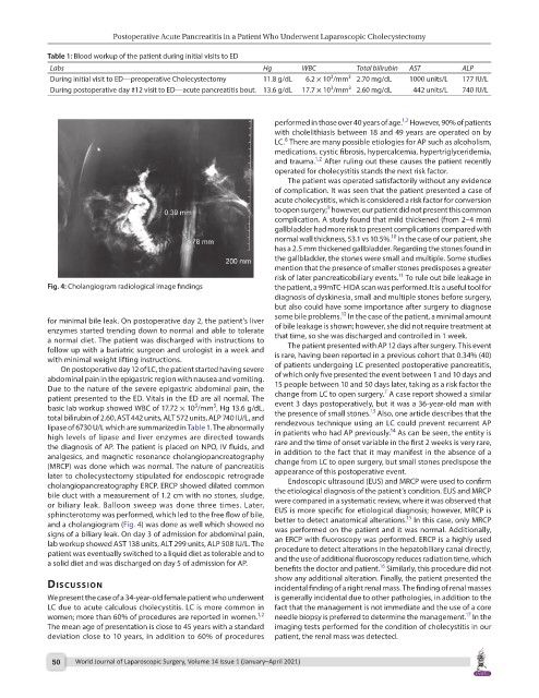
Fig. 4: Cholangiogram radiological image findings the patient, a 99mTC-HIDA scan was performed. It is a useful tool for
diagnosis of dyskinesia, small and multiple stones before surgery,
but also could have some importance after surgery to diagnose
12
some bile problems. In the case of the patient, a minimal amount
for minimal bile leak. On postoperative day 2, the patient’s liver
enzymes started trending down to normal and able to tolerate of bile leakage is shown; however, she did not require treatment at
a normal diet. The patient was discharged with instructions to that time, so she was discharged and controlled in 1 week.
follow up with a bariatric surgeon and urologist in a week and The patient presented with AP 12 days after surgery. This event
with minimal weight lifting instructions. is rare, having been reported in a previous cohort that 0.34% (40)
On postoperative day 12 of LC, the patient started having severe of patients undergoing LC presented postoperative pancreatitis,
abdominal pain in the epigastric region with nausea and vomiting. of which only five presented the event between 1 and 10 days and
Due to the nature of the severe epigastric abdominal pain, the 15 people between 10 and 50 days later, taking as a risk factor the
7
patient presented to the ED. Vitals in the ED are all normal. The change from LC to open surgery. A case report showed a similar
3
3
basic lab workup showed WBC of 17.72 × 10 /mm , Hg 13.6 g/dL, event 3 days postoperatively, but it was a 36-year-old man with
13
total bilirubin of 2.60, AST 442 units, ALT 572 units, ALP 740 IU/L, and the presence of small stones. Also, one article describes that the
lipase of 6730 U/L which are summarized in Table 1. The abnormally rendezvous technique using an LC could prevent recurrent AP
14
high levels of lipase and liver enzymes are directed towards in patients who had AP previously. As can be seen, the entity is
the diagnosis of AP. The patient is placed on NPO, IV fluids, and rare and the time of onset variable in the first 2 weeks is very rare,
analgesics, and magnetic resonance cholangiopancreatography in addition to the fact that it may manifest in the absence of a
(MRCP) was done which was normal. The nature of pancreatitis change from LC to open surgery, but small stones predispose the
later to cholecystectomy stipulated for endoscopic retrograde appearance of this postoperative event.
cholangiopancreatography ERCP. ERCP showed dilated common Endoscopic ultrasound (EUS) and MRCP were used to confirm
bile duct with a measurement of 1.2 cm with no stones, sludge, the etiological diagnosis of the patient’s condition. EUS and MRCP
or biliary leak. Balloon sweep was done three times. Later, were compared in a systematic review, where it was observed that
sphincterotomy was performed, which led to the free flow of bile, EUS is more specific for etiological diagnosis; however, MRCP is
15
and a cholangiogram (Fig. 4) was done as well which showed no better to detect anatomical alterations. In this case, only MRCP
signs of a biliary leak. On day 3 of admission for abdominal pain, was performed on the patient and it was normal. Additionally,
lab workup showed AST 138 units, ALT 299 units, ALP 508 IU/L. The an ERCP with fluoroscopy was performed. ERCP is a highly used
patient was eventually switched to a liquid diet as tolerable and to procedure to detect alterations in the hepatobiliary canal directly,
a solid diet and was discharged on day 5 of admission for AP. and the use of additional fluoroscopy reduces radiation time, which
16
benefits the doctor and patient. Similarly, this procedure did not
show any additional alteration. Finally, the patient presented the
dIscussIon incidental finding of a right renal mass. The finding of renal masses
We present the case of a 34-year-old female patient who underwent is generally incidental due to other pathologies, in addition to the
LC due to acute calculous cholecystitis. LC is more common in fact that the management is not immediate and the use of a core
1,2
17
women; more than 60% of procedures are reported in women. needle biopsy is preferred to determine the management. In the
The mean age of presentation is close to 45 years with a standard imaging tests performed for the condition of cholecystitis in our
deviation close to 10 years, in addition to 60% of procedures patient, the renal mass was detected.
50 World Journal of Laparoscopic Surgery, Volume 14 Issue 1 (January–April 2021)