
Page 29 - WJOLS - Laparoscopic Journal
P. 29
Review of Literatures on Laparoscopic Prosthetic Repair of Giant Hiatal Hernia than Pure Anatomical Repair of Crura
Tension-free Techniques
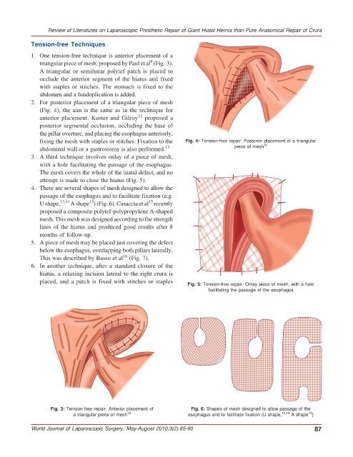
1. One tension-free technique is anterior placement of a
9
triangular piece of mesh, proposed by Paul et al (Fig. 3).
A triangular or semilunar polytef patch is placed to
occlude the anterior segment of the hiatus and fixed
with staples or stitches. The stomach is fixed to the
abdomen and a fundoplication is added.
2. For posterior placement of a triangular piece of mesh
(Fig. 4), the aim is the same as in the technique for
11
anterior placement. Kuster and Gilroy proposed a
posterior segmental occlusion, occluding the base of
the pillar overture, and placing the esophagus anteriorly,
fixing the mesh with staples or stitches. Fixation to the Fig. 4: Tension-free repair: Posterior placement of a triangular
11
abdominal wall or a gastrostomy is also performed. 12 piece of mesh
3. A third technique involves onlay of a piece of mesh,
with a hole facilitating the passage of the esophagus.
The mesh covers the whole of the hiatal defect, and no
attempt is made to close the hiatus (Fig. 5).
4. There are several shapes of mesh designed to allow the
passage of the esophagus and to facilitate fixation (e.g.
15
15
U shape, 13,14 A shape ) (Fig. 6). Casaccia et al recently
proposed a composite polytef-polypropylene A-shaped
mesh. This mesh was designed according to the strength
lines of the hiatus and produced good results after 8
months of follow-up.
5. A piece of mesh may be placed just covering the defect
below the esophagus, overlapping both pillars laterally.
16
This was described by Basso et al (Fig. 7).
6. In another technique, after a standard closure of the
hiatus, a relaxing incision lateral to the right crura is
placed, and a patch is fixed with stitches or staples
Fig. 5: Tension-free repair: Onlay piece of mesh, with a hole
facilitating the passage of the esophagus
Fig. 3: Tension-free repair: Anterior placement of Fig. 6: Shapes of mesh designed to allow passage of the
15
a triangular piece of mesh 10 esophagus and to facilitate fixation (U shape, 13,14 A shape )
World Journal of Laparoscopic Surgery, May-August 2010;3(2):85-90 87