
Page 48 - WJOLS - Laparoscopic Journal
P. 48
WJOLS
Minimally Invasive Esophagectomy (MIE): Techniques and Outcomes
STEPS OF THREE-STAGE ESOPHAGECTOMY esophagus is lifted and cut. The cut is extended upward to
the root of the neck. The vagus nerve is identified, and the
Stage 1: Thoracoscopic Esophageal
Mobilization vagal fibers going to the bronchus are preserved.
The dissection is started posteriorly between the
General anesthesia with single lung ventilation is used. The esophagus and vertebrae. All the fibro fatty tissues together
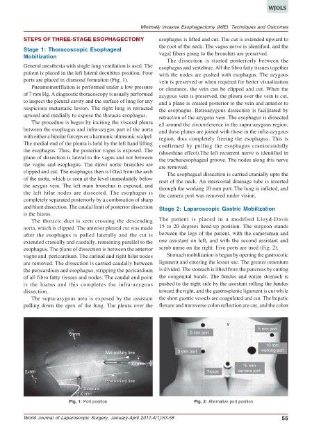
patient is placed in the left lateral decubitus position. Four with the nodes are pushed with esophagus. The azygous
ports are placed in diamond formation (Fig. 1). vein is preserved or when required for better visualization
Pneumoinsufflation is performed under a low pressure or clearance, the vein can be clipped and cut. When the
of 7 mm Hg. A diagnostic thoracoscopy is usually performed azygous vein is preserved, the pleura over the vein is cut,
to inspect the pleural cavity and the surface of lung for any and a plane is created posterior to the vein and anterior to
suspicious metastatic lesion. The right lung is retracted the esophagus. Retroazygous dissection is facilitated by
upward and medially to expose the thoracic esophagus. retraction of the azygous vein. The esophagus is dissected
The procedure is begun by incising the visceral pleura all around the circumference in the supra-azygous region,
between the esophagus and infra-azygos part of the aorta and these planes are joined with those in the infra-azygous
with either a bipolar forceps or a harmonic ultrasonic scalpel. region, thus completely freeing the esophagus. This is
The medial end of the pleura is held by the left hand lifting confirmed by pulling the esophagus craniocaudally
the esophagus. Thus, the posterior vagus is exposed. The (shoeshine effect).The left recurrent nerve is identified in
plane of dissection is lateral to the vagus and not between the tracheoesophageal groove. The nodes along this nerve
the vagus and esophagus. The direct aortic branches are are removed.
clipped and cut. The esophagus then is lifted from the arch The esophageal dissection is carried cranially upto the
of the aorta, which is seen at the level immediately below root of the neck. An intercostal drainage tube is inserted
the azygos vein. The left main bronchus is exposed, and through the working 10 mm port. The lung is inflated, and
the left hilar nodes are dissected. The esophagus is the camera port was removed under vision.
completely separated posteriorly by a combination of sharp
and blunt dissection. The caudal limit of posterior dissection Stage 2: Laparoscopic Gastric Mobilization
is the hiatus.
The thoracic duct is seen crossing the descending The patient is placed in a modified Lloyd-Davis
aorta, which is clipped. The anterior pleural cut was made 15 to 20 degrees head-up position. The surgeon stands
after the esophagus is pulled laterally and the cut is between the legs of the patient, with the cameraman and
extended cranially and caudally, remaining parallel to the one assistant on left, and with the second assistant and
esophagus. The plane of dissection is between the anterior scrub nurse on the right. Five ports are used (Fig. 2).
vagus and pericardium. The carinal and right hilar nodes Stomach mobilization is begun by opening the gastrocolic
are removed. The dissection is carried caudally between ligament and entering the lesser sac. The greater omentum
the pericardium and esophagus, stripping the pericardium is divided. The stomach is lifted from the pancreas by cutting
of all fibro fatty tissues and nodes. The caudal end point the congenital bands. The fundus and entire stomach is
is the hiatus and this completes the infra-azygous pushed to the right side by the assistant rolling the fundus
dissection. toward the right, and the gastrosplenic ligament is cut while
The supra-azygous area is exposed by the assistant the short gastric vessels are coagulated and cut. The hepatic
pulling down the apex of the lung. The pleura over the flexure and transverse colon reflection are cut, and the colon
Fig. 1: Port position Fig. 2: Alternative port position
World Journal of Laparoscopic Surgery, January-April 2011;4(1):53-58 55