Operative Steps of Laparoscopic Appendectomy
Patient Position
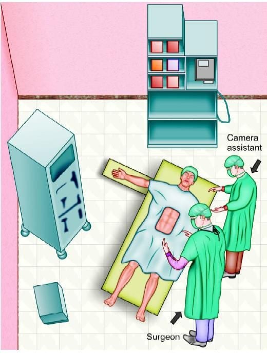
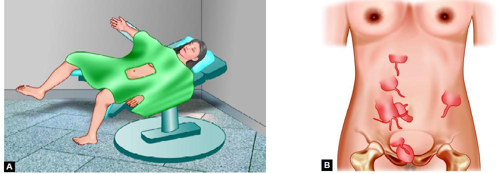
The patient is in a supine position, arms tucked at the side. The surgeon stands on the left side of the patient with the camera holder-assistant. For maintaining a coaxial alignment surgeon should stand near the left shoulder and the monitor should be placed near the right hip facing towards the surgeon. In a female, the lithotomic position should be used because it may be necessary to use a uterine manipulator in difficult diagnosis.

Patient position and set-up of the operating team

(A) Patient position in the female; (B) Variation in the position of the appendix
Port Position
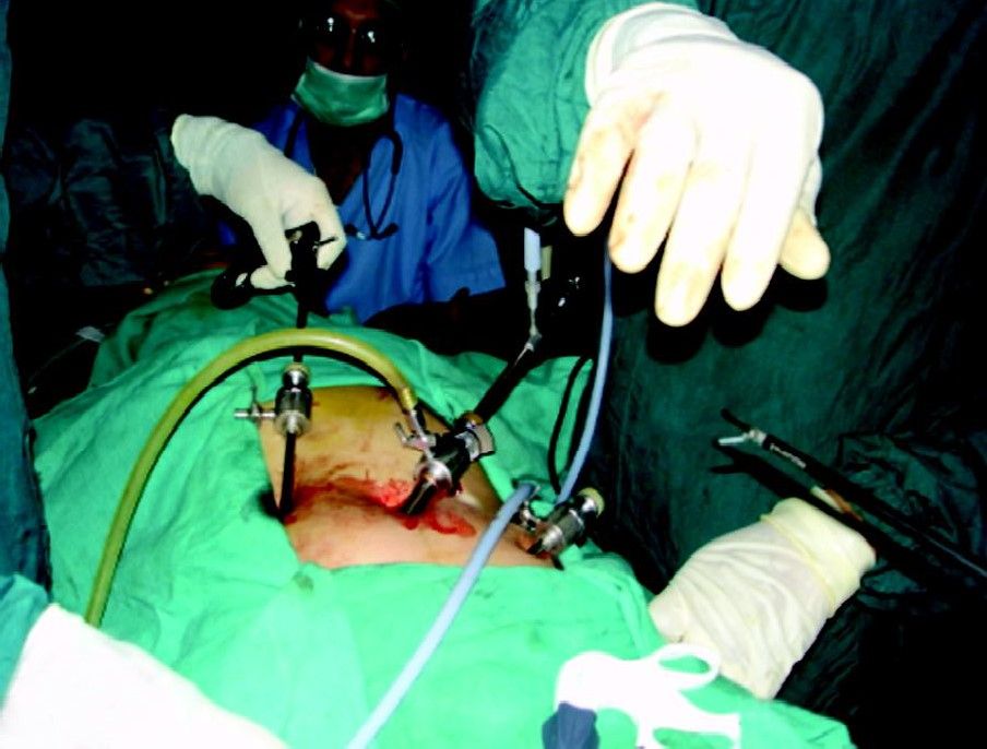
• Total 3 trocar should be used
• Two 10 mm, umbilical and left lower quadrant trocar
• One 5 mm right upper quadrant trocar
• The right upper quadrant trocar can be moved below the bikini line in females.

Port position in appendicectomy
Alternative Port and Theater Set-up
In beauty conscious females for a cosmetic reason, the baseball diamond concept of port position can be altered and three ports should be placed in such a way so that the two 5 mm port will be below the bikini line. Access should be performed by a 10 mm umbilical port. Once the telescope is inside one 5 mm port should be placed in left iliac fossa below the bikini line under vision. The second 5 mm port should be placed in the right iliac fossa, just mirror image of left port. After fixing all the ports in position, one another 5 mm telescope is introduced through left iliac fossa, and surgery should be performed through umbilical port (for right hand) and left iliac fossa port (for left hand). In this alternative port position, a 60° manipulation angle cannot be achieved and it is ergonomically difficult for the surgeon, but the patient will get a cosmetic benefit. This alternative port position for laparoscopic appendicectomy should not be performed in the case of retrocecal appendix or perforated appendix. Alternative port position in a beauty-conscious female.

Alternative port position
Operative Technique
Pneumoperitoneum is created in the usual fashion. Three ports are used in atraumatic grasper (Endo Babcock or Dolphin Nose Grasper) and are inserted via the right upper quadrant trocar. The cecum is retracted upward toward the liver. In most cases, this maneuver will elevate the appendix in the optical field of the telescope.
Retraction of Appendix
The appendix is grasped at its tip with a 5 mm claw grasper via the RUQ trocar. It is held in an upward position. After the pelvis is inspected the appendix is identified, mobilized, and examined properly. Periappendiceal or pericecal adhesion is lysed using either bipolar or harmonics and scissors. The left lower quadrant (LLQ) grasper is used to create a mesenteric window behind the base of the appendix.

Retraction of an appendix

Retraction of appendix and creation of a window over mesoappendix
A dolphin nose grasper is used to create a mesenteric window under the base of the appendix. The window should be made as close as possible to the base of the appendix and should be approximately 1 cm in size. The mesoappendix may be coagulated using bipolar or harmonics, it can be or clipped or stapled. It can also be sutured and cut with laparoscopic scissors to skeletonize the appendix.
Extracorporeal knotting performed (Meltzer or Tayside knot) for mesoappendix as well as an appendix. Two endoloop sutures are passed sequentially through one of the 5 mm ports and pushed around the base of an appendix on top of each other at a distance of 3 to 5 mm. A third endoloop suture can be applied 6 mm distal to the second suture so that surgeon will cut between second and third. Hulka tubal clips used for tubal sterilization can also be used sometimes to secure the proximal or distal portion of the appendix. The luminal portion of the appendiceal stump is sterilized with electrosurgery to prevent spillage and contamination of the peritoneal cavity. Betadine can be applied over the stump of the appendix and thorough suction and irrigation are performed either by normal saline or Ringer’s lactate solution. After extracting the appendix out of the abdominal cavity surgeon should examine the abdomen for any possible bowel injury or hemorrhage.
Patient Position
The patient is in a supine position, arms tucked at the side. The surgeon stands on the left side of the patient with the camera holder-assistant. For maintaining a coaxial alignment surgeon should stand near the left shoulder and the monitor should be placed near the right hip facing towards the surgeon. In a female, the lithotomic position should be used because it may be necessary to use a uterine manipulator in difficult diagnosis.

Patient position and set-up of the operating team

(A) Patient position in the female; (B) Variation in the position of the appendix
Port Position
• Total 3 trocar should be used
• Two 10 mm, umbilical and left lower quadrant trocar
• One 5 mm right upper quadrant trocar
• The right upper quadrant trocar can be moved below the bikini line in females.

Port position in appendicectomy
Alternative Port and Theater Set-up
In beauty conscious females for a cosmetic reason, the baseball diamond concept of port position can be altered and three ports should be placed in such a way so that the two 5 mm port will be below the bikini line. Access should be performed by a 10 mm umbilical port. Once the telescope is inside one 5 mm port should be placed in left iliac fossa below the bikini line under vision. The second 5 mm port should be placed in the right iliac fossa, just mirror image of left port. After fixing all the ports in position, one another 5 mm telescope is introduced through left iliac fossa, and surgery should be performed through umbilical port (for right hand) and left iliac fossa port (for left hand). In this alternative port position, a 60° manipulation angle cannot be achieved and it is ergonomically difficult for the surgeon, but the patient will get a cosmetic benefit. This alternative port position for laparoscopic appendicectomy should not be performed in the case of retrocecal appendix or perforated appendix. Alternative port position in a beauty-conscious female.

Alternative port position
Operative Technique
Pneumoperitoneum is created in the usual fashion. Three ports are used in atraumatic grasper (Endo Babcock or Dolphin Nose Grasper) and are inserted via the right upper quadrant trocar. The cecum is retracted upward toward the liver. In most cases, this maneuver will elevate the appendix in the optical field of the telescope.
Retraction of Appendix
The appendix is grasped at its tip with a 5 mm claw grasper via the RUQ trocar. It is held in an upward position. After the pelvis is inspected the appendix is identified, mobilized, and examined properly. Periappendiceal or pericecal adhesion is lysed using either bipolar or harmonics and scissors. The left lower quadrant (LLQ) grasper is used to create a mesenteric window behind the base of the appendix.

Retraction of an appendix

Retraction of appendix and creation of a window over mesoappendix
A dolphin nose grasper is used to create a mesenteric window under the base of the appendix. The window should be made as close as possible to the base of the appendix and should be approximately 1 cm in size. The mesoappendix may be coagulated using bipolar or harmonics, it can be or clipped or stapled. It can also be sutured and cut with laparoscopic scissors to skeletonize the appendix.
Extracorporeal knotting performed (Meltzer or Tayside knot) for mesoappendix as well as an appendix. Two endoloop sutures are passed sequentially through one of the 5 mm ports and pushed around the base of an appendix on top of each other at a distance of 3 to 5 mm. A third endoloop suture can be applied 6 mm distal to the second suture so that surgeon will cut between second and third. Hulka tubal clips used for tubal sterilization can also be used sometimes to secure the proximal or distal portion of the appendix. The luminal portion of the appendiceal stump is sterilized with electrosurgery to prevent spillage and contamination of the peritoneal cavity. Betadine can be applied over the stump of the appendix and thorough suction and irrigation are performed either by normal saline or Ringer’s lactate solution. After extracting the appendix out of the abdominal cavity surgeon should examine the abdomen for any possible bowel injury or hemorrhage.


