Laparoscopic Sigmoidectomy for Diverticular Disease
Outcomes after laparoscopic sigmoidectomy for diverticulitis are similar or even better to those seen in the open method, with faster recovery and decreased postoperative pain. Hand-assisted laparoscopic sigmoid resection for diverticulitis is also an attractive alternative to a "pure" laparoscopic method in complicated cases. The vascular approach for patients with benign diseases of the sigmoid colon is performed with the following steps.
Peritoneal Incision
The peritoneal incision can be similar to the cancer technique particularly in difficult cases (obesity, inflammatory mesocolon). In most cases, the surgeon should try to preserve the vascularization of the rectum and the left colic vessels. The opening of the peritoneum can be limited to the mesosigmoid parallel to the colon at mid-distance between the colon and the root of the mesosigmoid. An initial lateral mobilization of the sigmoid can be useful in this approach. The branches of the sigmoid arterial trunk can be divided separately anteriorly to inferior mesenteric vessels or together after creating windows in the mesentery to divide the various branches. A linear stapler or, better, the LigaSure Atlas 10 mm device can be used for this task.
Resection of the Specimen
In diverticular disease, one should perform the distal resection of the bowel below the rectosigmoid junction. The rectosigmoid junction is located just above the peritoneal reflexion, at the pouch of Douglas. It is preferred to perform the mobilization of the splenic flexure at this moment, before resection at the proximal limit, using the same principles as described above.
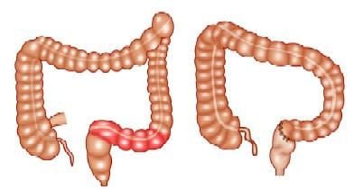
Before and after sigmoidectomy
Extraction of the Specimen
Before extracting the colon, it is important to divide the mesocolon at the level of the proximal site of the division. After adequate mobilization is achieved, the colon is extracted through a suprapubic incision, protected by the plastic drape described above, and proximal division performed externally on a compliant and well-vascularized part of the colon. The anastomosis is performed as described above for cancer.
Special Considerations
Ureteral injuries are one of the most important complications, which can be avoided by a perfect exposure and the respect of the correct plane of dissection. Indeed, a dissection properly performed above the Toldt's fascia does not expose the ureter to accidental injury. Difficult cases, such as important inflammatory reaction, cancer invasion or adhesions, and, sometimes, endometriosis, may alter the anatomy of the region and render the identification of the ureter troublesome. In these special cases, prevention of ureteral injury may be facilitated by the use of infrared wires inserted in ureteral stents. The infrared light is cold and safe for use in close contact with the ureteral tissue, and, on the other side, makes it easy to recognize the structure under the light of an adequate laparoscope.
Outcomes after laparoscopic sigmoidectomy for diverticulitis are similar or even better to those seen in the open method, with faster recovery and decreased postoperative pain. Hand-assisted laparoscopic sigmoid resection for diverticulitis is also an attractive alternative to a "pure" laparoscopic method in complicated cases. The vascular approach for patients with benign diseases of the sigmoid colon is performed with the following steps.
Peritoneal Incision
The peritoneal incision can be similar to the cancer technique particularly in difficult cases (obesity, inflammatory mesocolon). In most cases, the surgeon should try to preserve the vascularization of the rectum and the left colic vessels. The opening of the peritoneum can be limited to the mesosigmoid parallel to the colon at mid-distance between the colon and the root of the mesosigmoid. An initial lateral mobilization of the sigmoid can be useful in this approach. The branches of the sigmoid arterial trunk can be divided separately anteriorly to inferior mesenteric vessels or together after creating windows in the mesentery to divide the various branches. A linear stapler or, better, the LigaSure Atlas 10 mm device can be used for this task.
Resection of the Specimen
In diverticular disease, one should perform the distal resection of the bowel below the rectosigmoid junction. The rectosigmoid junction is located just above the peritoneal reflexion, at the pouch of Douglas. It is preferred to perform the mobilization of the splenic flexure at this moment, before resection at the proximal limit, using the same principles as described above.
Before and after sigmoidectomy
Extraction of the Specimen
Before extracting the colon, it is important to divide the mesocolon at the level of the proximal site of the division. After adequate mobilization is achieved, the colon is extracted through a suprapubic incision, protected by the plastic drape described above, and proximal division performed externally on a compliant and well-vascularized part of the colon. The anastomosis is performed as described above for cancer.
Special Considerations
Ureteral injuries are one of the most important complications, which can be avoided by a perfect exposure and the respect of the correct plane of dissection. Indeed, a dissection properly performed above the Toldt's fascia does not expose the ureter to accidental injury. Difficult cases, such as important inflammatory reaction, cancer invasion or adhesions, and, sometimes, endometriosis, may alter the anatomy of the region and render the identification of the ureter troublesome. In these special cases, prevention of ureteral injury may be facilitated by the use of infrared wires inserted in ureteral stents. The infrared light is cold and safe for use in close contact with the ureteral tissue, and, on the other side, makes it easy to recognize the structure under the light of an adequate laparoscope.


