Laparoscopic Myomectomy
Fibroids are a common uterine tumor affecting 20 to 25 percent of women. Fibroid develops from the benign transformation of a single smooth muscle cell. The growth of myoma is dependent on many factors. Increased estrogen stimulation alone or together with growth hormone or human placental lactogen appears to be the major growth regulator of fibroid. The severities of symptoms depend on the number of tumor, size, and location. Many times they cause abdominal pressure so, urinary frequency, abdominal pain, or constipation. One of the common findings is DUB due to altered blood flow through the uterus. The fibroid many times does not affect pregnancy. In women with menorrhagia, the hematocrit is used to assess the degree of anemia. Patients with large broad ligament fibroid may require an intravenous pyelogram to rule out any ureteral obstruction. For anemic patients, preoperative treatment with gonadotropin-releasing hormone may enable restoration of a normal hematocrit and decrease the size of myoma and thus reduce the risk of transfusion. Intraoperatively use of dilute vasopressin helps to minimize blood loss. Vertical uterine incision bleeds less than the transverse incision. Single, vertical, anterior, midline incisions are least likely to form adhesion. Although sutures predispose to adhesions, they are often necessary to close the uterine defect. If the endometrial cavity is entered due to large myoma a patient who subsequently becomes pregnant should undergo cesarean delivery. Preoperative GnRH agonist has been used by some gynecologists to decrease myomas and intraoperative blood loss. The risk of future uterine rupture is a major concern following myomectomy. The difficulty of adequately closing the layers laparoscopically and the use of electro-coagulation may contribute to the risk of uterine rupture. Uteroperitoneal fistulas may follow laparoscopic myomectomy because meticulous laparoscopic approximation of all layers is very difficult. The use of electrosurgery for hemostasis inside the uterine defect may also increase the risk of uteroperitoneal fistula formation. The chances of postoperative adhesion are also quite high in the case of laparoscopic myomectomy. A single uterine incision for removal of multiple leiomyomas and subserosal approximation of the uterine defect should be done. Laparoscopic-assisted myomectomy can reduce the chance of this complication. The suturing will be done outside so decreasing the operating time and secure layered suturing ensures uterus does not rupture in later pregnancy. Pelvic observation during the laparoscopy allows the diagnosis and treatment of any other disease like endometriosis or adhesion. The criteria for LAM are myoma greater than 5 cm, numerous myomas, requiring vigorous use of a morcellator, deep intramural myoma, and removal that require uterine repair with sutures.
Procedure
Pedunculated myomas are easiest to remove by just coagulating and cutting the stalk. Diluted vasopressin may be injected in the base of the stalk.
Intramural fibroid requires more manipulation so dilute vasopressin should be injected to multiple sites between the myometrium and the fibroid capsule. A vertical incision is made on the serosa overlying the leiomyomas using the monopolar electrode. The incision is extended until it reaches the capsule. The myometrium retracts as an incision is made, exposing the tumor. Two grasping tooth forceps hold the edges of the myometrium and the suction irrigator is used as a blunt probe to remove the covering of the leiomyomas from its capsule. The myoma crew should be inserted into the fibroid to apply traction while the suction irrigation instrument can be used as a blunt dissector. The CO2 laser is used to further dissect capsular attachment. Vessels are electrocoagulated before being cut. After complete myoma removal, the uterine defect is irrigated, bleeding points are identified and controlled with the open jaw of bipolar. If the fibroid is small and the patient does not want baby, the edges of the uterine defects are approximated by coagulating the myometrium without suturing and tube ligation is performed. If the defect is deep situated, the edges of the defect should be approximated by using 4-0 PDS. The repair mainly involves the serosal and subserosal layer or can be in one layer. Sutures are applied at a distance of 5 mm. After repair, thorough suction and irrigation should be performed. Some gynecologists use adhesive medical glues over the suture line to prevent adhesion. Even in the hand of an expert, the laparoscopic myomectomy is difficult. Tumble square knot is better to use if the edges are in tension. Dundee jamming knot with continuous suturing may be used if there is not much tension followed by Aberdeen's termination. Precise suturing of several layers is almost impossible laparoscopically.
Intraligamentous and broad ligament fibroid are difficult to remove due to the risk of injury to the ureter and uterine artery at the time of dissection. Following a thorough exposure of ureter and vessels and depending on the location of myoma, an incision is made on the anterior or posterior leaf of the broad ligament and the leiomyomas are slowly shelled as other subserosal or intramural fibroid.
Throughout the procedure the location of the ureter is monitored, bleeding points are controlled by bipolar. The broad ligament and peritoneum are not closed in cases of broad ligament Myoma. If postoperative bleeding is suspected, a drain should be left.
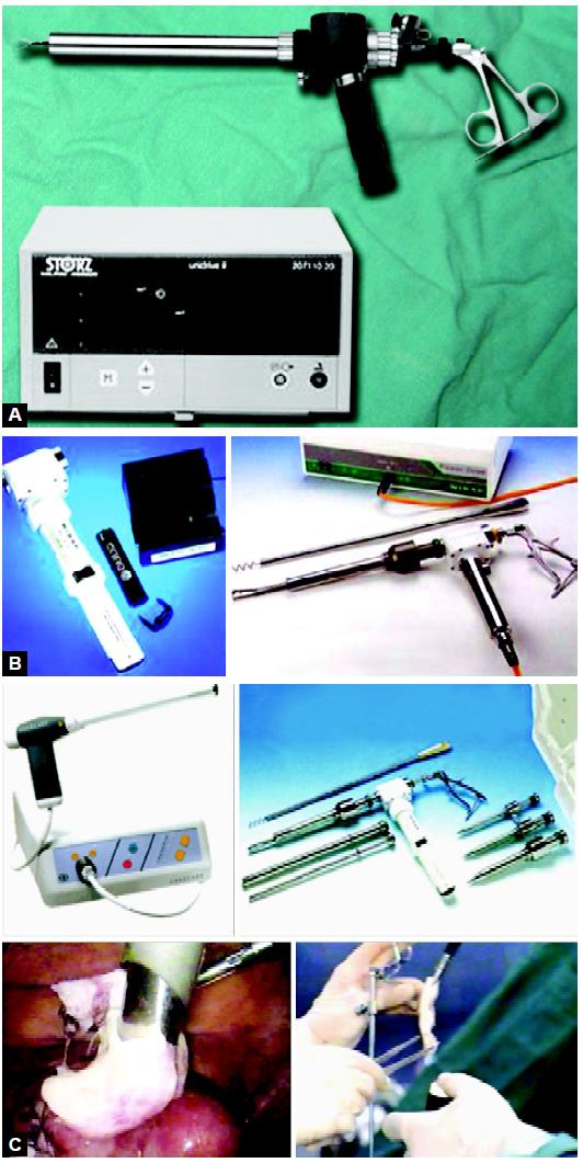
Morcellator is necessary equipment for myoma tissue retrieval
Removal of Myoma
Fibroid removal is one of the difficult and time-consuming procedures. Larger myoma may be removed through posterior colpotomy. In women with concurrent posterior cul-de-sac pathology, posterior colpotomy is not safe. Medium and large size fibroid is morcellated using a morcellator, scalpel or scissors. The process is ineffective for calcified myomas. For infected tissue and in the case of suspected carcinoma tissue retrieval bag should be used. Many sizes of disposable tissue retrieval bags are available and hard rim of these retrieval bags is easy to negotiate inside the abdominal cavity.
For large size gynecological tissue, the colpotomy route is good for retrieval. Colpotomy can be done laparoscopically with the help of heel of the hook. Counter pushing by other instruments is effective. Sponge over sponge holding forceps is inserted in posterior vaginal fornix by one assistant and the surgeon cuts the vaginal fascia between both the uterosacral ligaments with the heel of hook. The use of a morcellator is another way that facilitates the grinding of solid tissue and then these can be taken out without any difficulty. Recently many companies have launched a battery-operated morcellator. The morcellator is an important instrument for tissue retrieval in myomectomy and splenectomy.
Fibroids are a common uterine tumor affecting 20 to 25 percent of women. Fibroid develops from the benign transformation of a single smooth muscle cell. The growth of myoma is dependent on many factors. Increased estrogen stimulation alone or together with growth hormone or human placental lactogen appears to be the major growth regulator of fibroid. The severities of symptoms depend on the number of tumor, size, and location. Many times they cause abdominal pressure so, urinary frequency, abdominal pain, or constipation. One of the common findings is DUB due to altered blood flow through the uterus. The fibroid many times does not affect pregnancy. In women with menorrhagia, the hematocrit is used to assess the degree of anemia. Patients with large broad ligament fibroid may require an intravenous pyelogram to rule out any ureteral obstruction. For anemic patients, preoperative treatment with gonadotropin-releasing hormone may enable restoration of a normal hematocrit and decrease the size of myoma and thus reduce the risk of transfusion. Intraoperatively use of dilute vasopressin helps to minimize blood loss. Vertical uterine incision bleeds less than the transverse incision. Single, vertical, anterior, midline incisions are least likely to form adhesion. Although sutures predispose to adhesions, they are often necessary to close the uterine defect. If the endometrial cavity is entered due to large myoma a patient who subsequently becomes pregnant should undergo cesarean delivery. Preoperative GnRH agonist has been used by some gynecologists to decrease myomas and intraoperative blood loss. The risk of future uterine rupture is a major concern following myomectomy. The difficulty of adequately closing the layers laparoscopically and the use of electro-coagulation may contribute to the risk of uterine rupture. Uteroperitoneal fistulas may follow laparoscopic myomectomy because meticulous laparoscopic approximation of all layers is very difficult. The use of electrosurgery for hemostasis inside the uterine defect may also increase the risk of uteroperitoneal fistula formation. The chances of postoperative adhesion are also quite high in the case of laparoscopic myomectomy. A single uterine incision for removal of multiple leiomyomas and subserosal approximation of the uterine defect should be done. Laparoscopic-assisted myomectomy can reduce the chance of this complication. The suturing will be done outside so decreasing the operating time and secure layered suturing ensures uterus does not rupture in later pregnancy. Pelvic observation during the laparoscopy allows the diagnosis and treatment of any other disease like endometriosis or adhesion. The criteria for LAM are myoma greater than 5 cm, numerous myomas, requiring vigorous use of a morcellator, deep intramural myoma, and removal that require uterine repair with sutures.
Procedure
Pedunculated myomas are easiest to remove by just coagulating and cutting the stalk. Diluted vasopressin may be injected in the base of the stalk.
Intramural fibroid requires more manipulation so dilute vasopressin should be injected to multiple sites between the myometrium and the fibroid capsule. A vertical incision is made on the serosa overlying the leiomyomas using the monopolar electrode. The incision is extended until it reaches the capsule. The myometrium retracts as an incision is made, exposing the tumor. Two grasping tooth forceps hold the edges of the myometrium and the suction irrigator is used as a blunt probe to remove the covering of the leiomyomas from its capsule. The myoma crew should be inserted into the fibroid to apply traction while the suction irrigation instrument can be used as a blunt dissector. The CO2 laser is used to further dissect capsular attachment. Vessels are electrocoagulated before being cut. After complete myoma removal, the uterine defect is irrigated, bleeding points are identified and controlled with the open jaw of bipolar. If the fibroid is small and the patient does not want baby, the edges of the uterine defects are approximated by coagulating the myometrium without suturing and tube ligation is performed. If the defect is deep situated, the edges of the defect should be approximated by using 4-0 PDS. The repair mainly involves the serosal and subserosal layer or can be in one layer. Sutures are applied at a distance of 5 mm. After repair, thorough suction and irrigation should be performed. Some gynecologists use adhesive medical glues over the suture line to prevent adhesion. Even in the hand of an expert, the laparoscopic myomectomy is difficult. Tumble square knot is better to use if the edges are in tension. Dundee jamming knot with continuous suturing may be used if there is not much tension followed by Aberdeen's termination. Precise suturing of several layers is almost impossible laparoscopically.
Intraligamentous and broad ligament fibroid are difficult to remove due to the risk of injury to the ureter and uterine artery at the time of dissection. Following a thorough exposure of ureter and vessels and depending on the location of myoma, an incision is made on the anterior or posterior leaf of the broad ligament and the leiomyomas are slowly shelled as other subserosal or intramural fibroid.
Throughout the procedure the location of the ureter is monitored, bleeding points are controlled by bipolar. The broad ligament and peritoneum are not closed in cases of broad ligament Myoma. If postoperative bleeding is suspected, a drain should be left.
Morcellator is necessary equipment for myoma tissue retrieval
Removal of Myoma
Fibroid removal is one of the difficult and time-consuming procedures. Larger myoma may be removed through posterior colpotomy. In women with concurrent posterior cul-de-sac pathology, posterior colpotomy is not safe. Medium and large size fibroid is morcellated using a morcellator, scalpel or scissors. The process is ineffective for calcified myomas. For infected tissue and in the case of suspected carcinoma tissue retrieval bag should be used. Many sizes of disposable tissue retrieval bags are available and hard rim of these retrieval bags is easy to negotiate inside the abdominal cavity.
For large size gynecological tissue, the colpotomy route is good for retrieval. Colpotomy can be done laparoscopically with the help of heel of the hook. Counter pushing by other instruments is effective. Sponge over sponge holding forceps is inserted in posterior vaginal fornix by one assistant and the surgeon cuts the vaginal fascia between both the uterosacral ligaments with the heel of hook. The use of a morcellator is another way that facilitates the grinding of solid tissue and then these can be taken out without any difficulty. Recently many companies have launched a battery-operated morcellator. The morcellator is an important instrument for tissue retrieval in myomectomy and splenectomy.


