Laparoscopic Anatomy of Uterus
Benign uterine diseases of the uterus are very common and need hysterectomy and laparotomy. Most of these diseases can be performed laparoscopically. Laparoscopic-assisted vaginal hysterectomy is increasingly becoming popular. Many women come to the doctor and say they want a “laser” hysterectomy. What they usually mean is a laparoscopically assisted vaginal hysterectomy or LAVH. Laparoscopically assisted vaginal hysterectomy (LAVH) is a procedure using laparoscopic surgical techniques and instruments to remove the uterus and/or tubes and ovaries through the vagina. The technique used to use lasers but now lasers have been mostly replaced by surgical clips, cautery, or suturing. First laparoscopic hysterectomy was done by Reich et al in 1989. It is a technique made to replace abdominal hysterectomy.
The normal nulliparous uterus is approximately 8 cm in length and angled forward so the fundus lies over the posterior surface of the bladder. The uterus is all around covered with peritoneum except where the bladder touches the lower uterine segment at the anterior cul-de-sac and laterally at the broad ligament. Two important arteries, uterine and ovarian are of great significance in uterine surgery. The uterine arise from the internal iliac. They pass medially on the levator ani muscle, cross the ureter, and ultimately divide into ascending and descending branches. The uterine artery runs in a tortuous course within the broad ligaments. The uterine arteries ascending branch terminates by anatomizing with the ovarian artery. From anterior to posterior, the following important tubular structures are found crossing the brim of the true pelvis: The round ligament of the uterus, the infundibulopelvic ligament, which contains the gonadal vessels and the ureter. The ovaries and fallopian tubes are found between the round ligament and the infundibulopelvic ligament.

Anatomy of the uterus. (1) Umbilical artery; (2) Ureter; (3) Uterine artery; (4) Internal iliac artery; (5) Ovarian artery; (6) Common iliac artery; (7) Uterosacral ligament

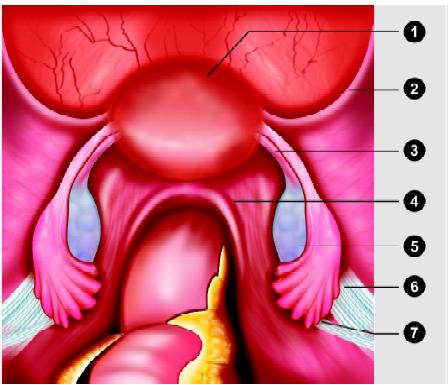
Position of uterus. (1) Uterus; (2) Round ligament; (3) Utero- ovarian ligament ( proper ovarian l igament); (4) Uterosacral ligament; (5) Ovary; (6) Suspensory ligament of the ovary; (7) Ureter
The ovarian ligaments run from the ovaries to the lateral border of the uterus. The ovary is attached to the pelvic sidewall with infundibulopelvic ligament, which carries the ovarian artery. One of the common mistakes is the injury of the ureter during dissection of the infundibulopelvic ligament. If the uterus deviates to the contralateral side with the help of uterine manipulator infundibulopelvic ligament is spread out and a pelvic sidewall triangle is created. The base of this triangle is the round ligament, the medial side is the infundibulopelvic ligament, and the lateral side is the external iliac artery. The apex of this triangle is the point at which the infundibulopelvic ligament crosses the external iliac artery. The ureter always enters medially to this triangle into the pelvis. It is visible under the peritoneum overlying the external iliac artery.
The ureters enter the pelvis in close proximity to the female pelvic organ and are at risk for injury during laparoscopic surgery of these organs. As the ureter course medially over the bifurcation of the iliac vessels, they pass obliquely under the ovarian vessels and then run in close proximity to the uterine artery. Laparoscopy hysterectomy needs careful identification of ureter with some dissection of retroperitoneum. An incision is made in the peritoneum overlying the pelvic sidewall triangle between the fallopian tube and the iliac vessel. Pelvic lymph node dissection is also necessary if gynecologist plan to perform a radical laparoscopic hysterectomy. Node dissection as far as distal as Cloquet’s node in the femoral triangle may be included and proximally dissection may be necessary up to para-aortic lymph node.
Benign uterine diseases of the uterus are very common and need hysterectomy and laparotomy. Most of these diseases can be performed laparoscopically. Laparoscopic-assisted vaginal hysterectomy is increasingly becoming popular. Many women come to the doctor and say they want a “laser” hysterectomy. What they usually mean is a laparoscopically assisted vaginal hysterectomy or LAVH. Laparoscopically assisted vaginal hysterectomy (LAVH) is a procedure using laparoscopic surgical techniques and instruments to remove the uterus and/or tubes and ovaries through the vagina. The technique used to use lasers but now lasers have been mostly replaced by surgical clips, cautery, or suturing. First laparoscopic hysterectomy was done by Reich et al in 1989. It is a technique made to replace abdominal hysterectomy.
The normal nulliparous uterus is approximately 8 cm in length and angled forward so the fundus lies over the posterior surface of the bladder. The uterus is all around covered with peritoneum except where the bladder touches the lower uterine segment at the anterior cul-de-sac and laterally at the broad ligament. Two important arteries, uterine and ovarian are of great significance in uterine surgery. The uterine arise from the internal iliac. They pass medially on the levator ani muscle, cross the ureter, and ultimately divide into ascending and descending branches. The uterine artery runs in a tortuous course within the broad ligaments. The uterine arteries ascending branch terminates by anatomizing with the ovarian artery. From anterior to posterior, the following important tubular structures are found crossing the brim of the true pelvis: The round ligament of the uterus, the infundibulopelvic ligament, which contains the gonadal vessels and the ureter. The ovaries and fallopian tubes are found between the round ligament and the infundibulopelvic ligament.

Anatomy of the uterus. (1) Umbilical artery; (2) Ureter; (3) Uterine artery; (4) Internal iliac artery; (5) Ovarian artery; (6) Common iliac artery; (7) Uterosacral ligament

Position of uterus. (1) Uterus; (2) Round ligament; (3) Utero- ovarian ligament ( proper ovarian l igament); (4) Uterosacral ligament; (5) Ovary; (6) Suspensory ligament of the ovary; (7) Ureter
The ovarian ligaments run from the ovaries to the lateral border of the uterus. The ovary is attached to the pelvic sidewall with infundibulopelvic ligament, which carries the ovarian artery. One of the common mistakes is the injury of the ureter during dissection of the infundibulopelvic ligament. If the uterus deviates to the contralateral side with the help of uterine manipulator infundibulopelvic ligament is spread out and a pelvic sidewall triangle is created. The base of this triangle is the round ligament, the medial side is the infundibulopelvic ligament, and the lateral side is the external iliac artery. The apex of this triangle is the point at which the infundibulopelvic ligament crosses the external iliac artery. The ureter always enters medially to this triangle into the pelvis. It is visible under the peritoneum overlying the external iliac artery.
The ureters enter the pelvis in close proximity to the female pelvic organ and are at risk for injury during laparoscopic surgery of these organs. As the ureter course medially over the bifurcation of the iliac vessels, they pass obliquely under the ovarian vessels and then run in close proximity to the uterine artery. Laparoscopy hysterectomy needs careful identification of ureter with some dissection of retroperitoneum. An incision is made in the peritoneum overlying the pelvic sidewall triangle between the fallopian tube and the iliac vessel. Pelvic lymph node dissection is also necessary if gynecologist plan to perform a radical laparoscopic hysterectomy. Node dissection as far as distal as Cloquet’s node in the femoral triangle may be included and proximally dissection may be necessary up to para-aortic lymph node.


