Totally Extraperitoneal Inguinal Hernia Repair
An inguinal hernia results from a hole or defect in the muscles, through which the peritoneum protrudes, forming the sac. Inguinal herniorrhaphy is one of the most common operations that general surgeons perform. Laparoscopic herniorrhaphy is being done at a time when laparoscopic cholecystectomy has shown definite benefits over the open technique. But unlike laparoscopic cholecystectomy, laparoscopic hernia repair is an advanced laparoscopic procedure and has a longer learning curve. In addition, TEP requires higher technical expertise for successful results.
Ger in 1982, attempted minimal access groin hernia repair by closing the opening of an indirect inguinal hernial sac using Michel clips. Bogojavlensky in 1989 modified the technique by intracorporeal suture of the deep ring after plugging a PPM into the sac. Toy and Smoot in 1991, described a technique of intraperitoneal Onlay mesh (IPOM) placement, where an intra-abdominal piece of polypropylene or e-PTFE was stapled over the myopectineal orifice without dissection of the peritoneum.
The present-day techniques of laparoscopic hernia repair evolved from Stoppa’s concept of pre-peritoneal reinforcement of fascia transversalis over the myopectineal orifice with its multiple openings by a prosthetic mesh. In the early 1990’ s Arregui and Doin described the transabdominal preperitoneal (TAPP) repair, where the abdominal cavity is first entered, peritoneum over the posterior wall of the inguinal canal is incised to enter into the avascular preperitoneal plane which is adequately dissected to place a large (15 × 10 cm) mesh over the hernial orifices. After the fixation of the mesh, the peritoneum is carefully sutured or stapled. TAPP approach has the advantage of identifying missed additional direct or femoral hernia during the first operation itself. Around the same time, Phillips and McKernan described the totally extraperitoneal (TEP) technique of endoscopic hernioplasty where the peritoneal cavity is not breached and the entire dissection is performed bluntly in the extraperitoneal space with a balloon device or the tip of the laparoscope itself. Advanced knowledge of the posterior anatomy of the inguinal region is imperative. Once the dissection is complete, a 15 × 10 cm mesh is stapled in place over the myopectineal orifice. It appears to be the most common endoscopic repair today. In both these repairs, the mesh in direct contact with the fascia of the transversalis muscle in the pre-peritoneal space, allows tissue ingrowths leading to the fixation of the mesh (as opposed to being in contact to the peritoneum as in IPOM repair where it is prone to migrate).
The technique of totally extraperitoneal (TEP) repair of inguinal hernia was described even before the TAPP technique; however, technical difficulties of working in closed space and anatomy with the limited working space hindered its popular acceptance. The effectiveness of this type of repair has been well established by the open operation of Stoppa.
Advantages of TEP
• Pneumoperitoneum is not required
• Less chance of dangerous vessel injury or bowel injury
• The view of the groin is better for dissection around the neck of the sac
• Continuity of peritoneum is not breached so it may not be closed.
Disadvantages of Preperitoneal Repair
• The identification of the correct plane of dissection is difficult
• The landmarks of hernia dissection can only be identified when they are encountered
• Reduction of the content of sac is difficult to ensure
• Sliding hernia is difficult to recognize from outside of the sac
• If the sac is cut it is difficult to close it again
• In recurrent hernia extensive adhesion make the dissection difficult because peritoneum may be adherent to the undersurface of scar
• There is always a chance of breach of peritoneum continuity and this will reduce the view
• Four ports generally are necessary for bilateral hernia surgery. Whereas, in TAPP only three ports are sufficient.
Preparation of the Patient
Preparation of the patient in totally preperitoneal hernia repair is the same as of the transabdominal hernia repair. Knowledge of the anatomy of the abdominal wall muscle and recognition of the transition zone that occurs at the arcuate line of Douglas is very important for totally preperitoneal hernia repair.
Approach to Preperitoneal Space
In the totally extraperitoneal repair of hernia, the main concern is to make an extraperitoneal space. The extraperitoneal space is made possible by the fact that the peritoneum in the suprapubic region can easily be separated from the anterior abdominal wall, thereby creating enough space for dissection.
A 2 cm longitudinal skin incision is made just below the umbilicus 1 cm lateral to the midline on the side of the hernia. The incision is deepened down to reach up to the anterior rectus sheath. All the subcutaneous fat is cleared and the rectus is opened under direct vision. Two- stay suture on each leaf of the rectus sheath is placed and the rectus muscle is retracted by two retractors downward towards symphysis pubis in an oblique fashion; we should never cross the posterior fascia of the rectus muscle while dissecting.

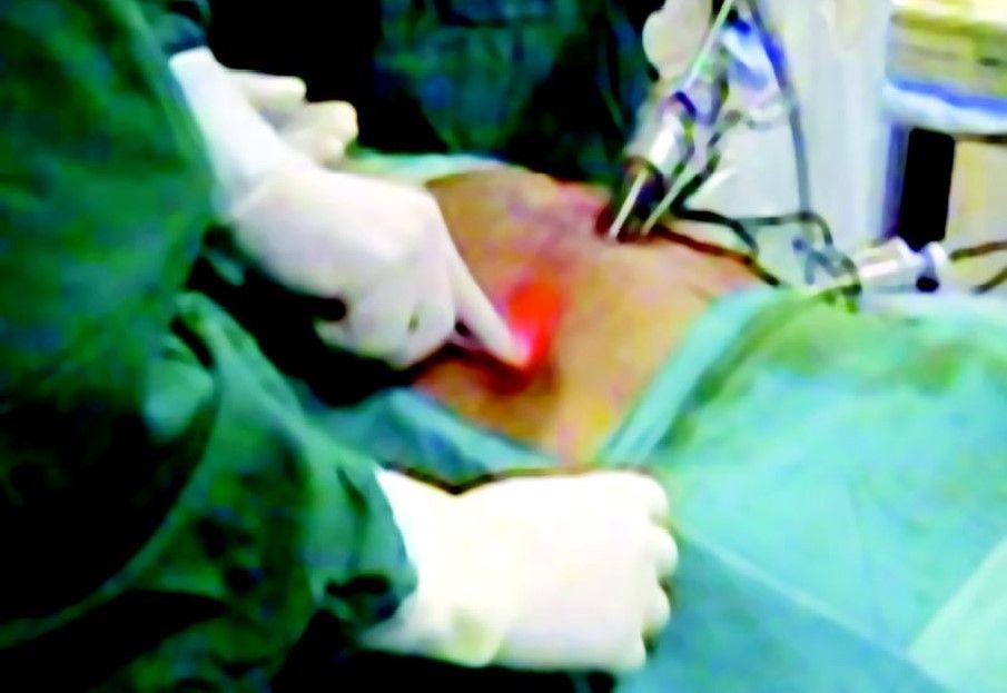
Access technique of totally extraperitoneal hernia repair
By finger or swab towards the hernia, dissection should perform carefully, preperitoneal space will be found below the arcuate line of Douglas.
Insertion of Port
A balloon dissector should be introduced with telescope and balloon is inflated for further dissection of the preperitoneal space. An 11 mm port is introduced without its sharp tip with a laparoscope at 30°. A small preperitoneal pocket is created by manipulating a laparoscope in a sweeping manner. If the balloon dissector is not available the glove finger can be tied around the suction irrigation instrument and can be used to create some preperitoneal space.

Making balloon dissection with the finger of gloves
Sweeping Movement of Telescope
Once the telescope is placed properly a 10 mm port is inserted under direct view approximately halfway between the symphysis pubis and the umbilicus. Another 5 mm port should be placed two fingers below and medial to the right anterior iliac spine. If the secondary port site is not seen clearly through the telescope one can infiltrate the port site with local anesthetic and look for the tip of the needle internally. This will ensure the exact placement of port and allow the tip of the trocar to be seen by a telescope at the time of insertion.
Introduction of the secondary port
Dissection of Preperitoneal Space and Cord Structures in TEP
In the totally extraperitoneal repair of hernia Stoppa's parietalization technique is used for dissection of the spermatic cord from the peritoneum by separating the elements of the spermatic cord from the peritoneum and peritoneal sac should be done. The dissection is started by tracing the inferior epigastric vessels towards the deep ring. The upper border of the hernia sac readily recognized because indirect hernia is lateral to the inferior epigastric vessels and direct hernia is medial to that. As the inguinal region is approached, the dissection is continued all around the sac to encircle the neck. The surgeon should try to remain close to peritoneum and dissection continues medially to separate vas from the sac. Under the neck of the sac, care should be taken to avoid injury of iliac vessels.

Dissection of preperitoneal space
In the case of direct inguinal hernia, the dissection is carried out from above downwards and progressed medially to the inferior epigastric vessels. The direct sac is freed from the transversalis fascia. Dissection should be continued until the peritoneum has reached the iliac vessels inferiorly. Care should be taken that any hole in peritoneum should not form otherwise it will be difficult to have good working space because the gas will escape into the abdominal cavity. If the hole is made anyway it should be identified and enlarged this will equalize the pressure on both sides of peritoneum and allows the peritoneum to drop back down due to gravity. A venting 5 mm port or Veress needle can be placed in the right upper quadrant at Palmer’s point to decompress the abdominal cavity.
The technique of insertion of mesh in the totally extraperitoneal repair of a hernia is the same as that of transabdominal preperitoneal. Mesh of the appropriate size usually 15 × 15 cm is used and rolled and loaded backward in one of the port. Mesh should be fixed by stapling first in its middle part three-finger above the superior limit of the internal ring. In totally extraperitoneal repair some surgeon does not use a staple, because peritoneum is not breached and once the gas from preperitoneal space is removed, it will place the mesh in its proper position. In 1 to 2 percent of cases of TEP conversion to open or TAPP may be necessary due to large peritoneal tear making the vision difficult or in the cases where content is not reduced completely.

Introduction of mesh

Placement of mesh
Ending of the Operation
At the end of the surgery, the abdomen should be examined for any possible bowel injury or hemorrhage. The entire instrument should be removed and then all the port. Generally, Vicryl is used for rectus and unabsorbable intradermal and stapler for skin. The adhesive sterile dressing should be applied over the wound.
An inguinal hernia results from a hole or defect in the muscles, through which the peritoneum protrudes, forming the sac. Inguinal herniorrhaphy is one of the most common operations that general surgeons perform. Laparoscopic herniorrhaphy is being done at a time when laparoscopic cholecystectomy has shown definite benefits over the open technique. But unlike laparoscopic cholecystectomy, laparoscopic hernia repair is an advanced laparoscopic procedure and has a longer learning curve. In addition, TEP requires higher technical expertise for successful results.
Ger in 1982, attempted minimal access groin hernia repair by closing the opening of an indirect inguinal hernial sac using Michel clips. Bogojavlensky in 1989 modified the technique by intracorporeal suture of the deep ring after plugging a PPM into the sac. Toy and Smoot in 1991, described a technique of intraperitoneal Onlay mesh (IPOM) placement, where an intra-abdominal piece of polypropylene or e-PTFE was stapled over the myopectineal orifice without dissection of the peritoneum.
The present-day techniques of laparoscopic hernia repair evolved from Stoppa’s concept of pre-peritoneal reinforcement of fascia transversalis over the myopectineal orifice with its multiple openings by a prosthetic mesh. In the early 1990’ s Arregui and Doin described the transabdominal preperitoneal (TAPP) repair, where the abdominal cavity is first entered, peritoneum over the posterior wall of the inguinal canal is incised to enter into the avascular preperitoneal plane which is adequately dissected to place a large (15 × 10 cm) mesh over the hernial orifices. After the fixation of the mesh, the peritoneum is carefully sutured or stapled. TAPP approach has the advantage of identifying missed additional direct or femoral hernia during the first operation itself. Around the same time, Phillips and McKernan described the totally extraperitoneal (TEP) technique of endoscopic hernioplasty where the peritoneal cavity is not breached and the entire dissection is performed bluntly in the extraperitoneal space with a balloon device or the tip of the laparoscope itself. Advanced knowledge of the posterior anatomy of the inguinal region is imperative. Once the dissection is complete, a 15 × 10 cm mesh is stapled in place over the myopectineal orifice. It appears to be the most common endoscopic repair today. In both these repairs, the mesh in direct contact with the fascia of the transversalis muscle in the pre-peritoneal space, allows tissue ingrowths leading to the fixation of the mesh (as opposed to being in contact to the peritoneum as in IPOM repair where it is prone to migrate).
The technique of totally extraperitoneal (TEP) repair of inguinal hernia was described even before the TAPP technique; however, technical difficulties of working in closed space and anatomy with the limited working space hindered its popular acceptance. The effectiveness of this type of repair has been well established by the open operation of Stoppa.
Advantages of TEP
• Pneumoperitoneum is not required
• Less chance of dangerous vessel injury or bowel injury
• The view of the groin is better for dissection around the neck of the sac
• Continuity of peritoneum is not breached so it may not be closed.
Disadvantages of Preperitoneal Repair
• The identification of the correct plane of dissection is difficult
• The landmarks of hernia dissection can only be identified when they are encountered
• Reduction of the content of sac is difficult to ensure
• Sliding hernia is difficult to recognize from outside of the sac
• If the sac is cut it is difficult to close it again
• In recurrent hernia extensive adhesion make the dissection difficult because peritoneum may be adherent to the undersurface of scar
• There is always a chance of breach of peritoneum continuity and this will reduce the view
• Four ports generally are necessary for bilateral hernia surgery. Whereas, in TAPP only three ports are sufficient.
Preparation of the Patient
Preparation of the patient in totally preperitoneal hernia repair is the same as of the transabdominal hernia repair. Knowledge of the anatomy of the abdominal wall muscle and recognition of the transition zone that occurs at the arcuate line of Douglas is very important for totally preperitoneal hernia repair.
Approach to Preperitoneal Space
In the totally extraperitoneal repair of hernia, the main concern is to make an extraperitoneal space. The extraperitoneal space is made possible by the fact that the peritoneum in the suprapubic region can easily be separated from the anterior abdominal wall, thereby creating enough space for dissection.
A 2 cm longitudinal skin incision is made just below the umbilicus 1 cm lateral to the midline on the side of the hernia. The incision is deepened down to reach up to the anterior rectus sheath. All the subcutaneous fat is cleared and the rectus is opened under direct vision. Two- stay suture on each leaf of the rectus sheath is placed and the rectus muscle is retracted by two retractors downward towards symphysis pubis in an oblique fashion; we should never cross the posterior fascia of the rectus muscle while dissecting.
Access technique of totally extraperitoneal hernia repair
By finger or swab towards the hernia, dissection should perform carefully, preperitoneal space will be found below the arcuate line of Douglas.
Insertion of Port
A balloon dissector should be introduced with telescope and balloon is inflated for further dissection of the preperitoneal space. An 11 mm port is introduced without its sharp tip with a laparoscope at 30°. A small preperitoneal pocket is created by manipulating a laparoscope in a sweeping manner. If the balloon dissector is not available the glove finger can be tied around the suction irrigation instrument and can be used to create some preperitoneal space.
Making balloon dissection with the finger of gloves
Sweeping Movement of Telescope
Once the telescope is placed properly a 10 mm port is inserted under direct view approximately halfway between the symphysis pubis and the umbilicus. Another 5 mm port should be placed two fingers below and medial to the right anterior iliac spine. If the secondary port site is not seen clearly through the telescope one can infiltrate the port site with local anesthetic and look for the tip of the needle internally. This will ensure the exact placement of port and allow the tip of the trocar to be seen by a telescope at the time of insertion.
Introduction of the secondary port
Dissection of Preperitoneal Space and Cord Structures in TEP
In the totally extraperitoneal repair of hernia Stoppa's parietalization technique is used for dissection of the spermatic cord from the peritoneum by separating the elements of the spermatic cord from the peritoneum and peritoneal sac should be done. The dissection is started by tracing the inferior epigastric vessels towards the deep ring. The upper border of the hernia sac readily recognized because indirect hernia is lateral to the inferior epigastric vessels and direct hernia is medial to that. As the inguinal region is approached, the dissection is continued all around the sac to encircle the neck. The surgeon should try to remain close to peritoneum and dissection continues medially to separate vas from the sac. Under the neck of the sac, care should be taken to avoid injury of iliac vessels.
Dissection of preperitoneal space
In the case of direct inguinal hernia, the dissection is carried out from above downwards and progressed medially to the inferior epigastric vessels. The direct sac is freed from the transversalis fascia. Dissection should be continued until the peritoneum has reached the iliac vessels inferiorly. Care should be taken that any hole in peritoneum should not form otherwise it will be difficult to have good working space because the gas will escape into the abdominal cavity. If the hole is made anyway it should be identified and enlarged this will equalize the pressure on both sides of peritoneum and allows the peritoneum to drop back down due to gravity. A venting 5 mm port or Veress needle can be placed in the right upper quadrant at Palmer’s point to decompress the abdominal cavity.
The technique of insertion of mesh in the totally extraperitoneal repair of a hernia is the same as that of transabdominal preperitoneal. Mesh of the appropriate size usually 15 × 15 cm is used and rolled and loaded backward in one of the port. Mesh should be fixed by stapling first in its middle part three-finger above the superior limit of the internal ring. In totally extraperitoneal repair some surgeon does not use a staple, because peritoneum is not breached and once the gas from preperitoneal space is removed, it will place the mesh in its proper position. In 1 to 2 percent of cases of TEP conversion to open or TAPP may be necessary due to large peritoneal tear making the vision difficult or in the cases where content is not reduced completely.
Introduction of mesh
Placement of mesh
Ending of the Operation
At the end of the surgery, the abdomen should be examined for any possible bowel injury or hemorrhage. The entire instrument should be removed and then all the port. Generally, Vicryl is used for rectus and unabsorbable intradermal and stapler for skin. The adhesive sterile dressing should be applied over the wound.

