Introduction
Heart valve surgery:
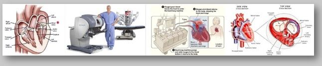
Heart valve surgery is applied to repair or replace disorders of heart valves. Blood which circulates between various chambers of the heart must flow through a heart valve. Blood that circulates out of heart into large arteries must flow through a heart valve. These valves open up enough so that blood can flow through. They then close, keeping blood from flowing backward.
There are four valves in heart:
- Aortic valve
- Mitral valve
- Tricuspid valve
- Pulmonary valve
In open surgery, the surgeon makes a large surgical cut in breastbone to reach the heart and aorta. Most people are connected to a heart-lung bypass machine or bypass pump. The heart is stopped while you are connected to this machine. Minimally invasive valve surgery is done through much smaller cuts than open surgery, or through a catheter inserted through the skin. Several different techniques are used:
- Laparoscopy or endoscopy
- Robot-assisted surgery
Thoracoscopic and Robotic management of Cardiac Valve Diseases
Robotic surgery is a approach in which a surgeon done surgery using a computer which remotely controls very small instruments attached to a robot. This method is done under general anesthesia. The surgeon sits at a computer station nearby and directs the movements of a robot. Small instruments are attached to the robot's arms. The surgeon first attached these instruments into your body through small surgical cuts. Under the surgeon's direction, the robot matches the doctor's hand movements to perform the procedure using the tiny instruments. A thin tube with a camera attached to the end of it endoscope allows the surgeon to view highly magnified three-dimensional images of your body on a monitor in real time.
Robotic surgery is a type of procedure that is similar to laparoscopic surgery. It also can be carried out through smaller surgical cuts than traditional open surgery. The small, precise movements that are possible with this type of surgery give it some advantages over standard endoscopic techniques. Sometimes robotic-assisted laparoscopy can allow a surgeon to perform a less-invasive procedure that was once only possible with more invasive open surgery. Once it is placed in the abdomen, a robotic arm is easier for the surgeon to use than the instruments in endoscopic surgery.
The robot reduces the surgeon's movements for example, moving 1/2 inch for every 1 inch the surgeon moves, which reduces some of the hand tremors and movements that might otherwise make the surgery less precise. Also, robotic instruments can access hard-to-reach areas of your body more easily through smaller surgical cuts compared to traditional open and laparoscopic surgery.
During robotic surgery, the surgeon can more easily see the area being operated on. The surgeon is also in a much more comfortable position and can move in a more natural way than during endoscopy. However, robotic surgery can take longer to perform, due to the amount of time needed to set up the robot. Also, the robot is expensive to use and may not be available in many hospitals.
Robotic surgery cannot be used for some complex procedures. For example, it is not appropriate for certain types of heart surgery that require greater ability to move instruments in the patient's chest. Because the surgical cuts are typically smaller than with traditional open surgery, robotic surgery may lead to:
- Faster recovery
- Less pain and bleeding
- Less risk of infection
- Shorter hospital stay
- Smaller scars
Thoracoscopic Cardiac Valve Surgery:
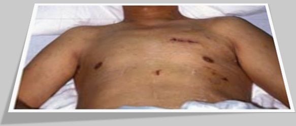
To begin the procedure, the patient is asleep and placed in the supine position, with a soft roll placed between the spine and the left scapula to elevate the left chest. Four tiny incisions of half an inch in length or less are made on each side of the chest. The pericardium, or fibrous sac enclosing the heart, is opened away from the phrenic nerve, which is responsible for innervating the diaphragm. In this, as in every maneuver of the operation, the thoracoscope provides clear, magnified visualization of all key anatomic structures. This technology enhances both the safety and precision of every maneuver. No computer-enhanced imaging is necessary, since all treated regions of the heart are seen in great detail with the thoracoscope.
Conclusion:
Thoracoscopic and Robotic management of Cardiac Valve Surgical procedures are considered to be even more minimally invasive than laparoscopic procedures, which are themselves a drastic improvement over open surgical techniques. State-of-the-art robotic and computer technology enables a surgeon to view the operating field through a special scope, utilizing fine hand movements that correspond with moments of robotic arms utilizing miniaturized equipment attached to the surgeon's wrist. The surgeon controls the movements of the robotic arms and instruments in magnified procedures regardless of operating area size.
World Laparoscopy Hospital, Cyber City, DLF Phase II, Gurugram, NCR Delhi, 122 002, India
PHONES:
For Training: +919811416838
For Treatment: +919811912768
For General Enquiry: +91(0)124 - 2351555


