Operative Technique of TVT-O
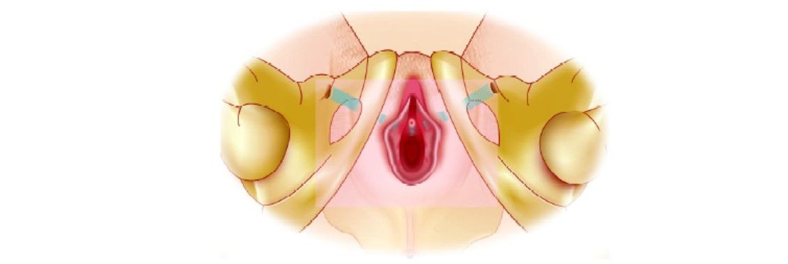
The patient should be positioned in dorsal lithotomy with hips hyperflexed with buttock flush to the edge of the table. Make the exit points by tracing a horizontal line at the level of the urethral meatus, and a second line parallel and 2 cm above the first line. Locate the exit points on the second line, 2 cm lateral of the folds of the thigh. Using Allis clamps for traction make a 1 cm midline vaginal incision starting 1 cm proximal to the urethral meatus.

Position the patient, mark thigh exit point and make a vaginal midline incision
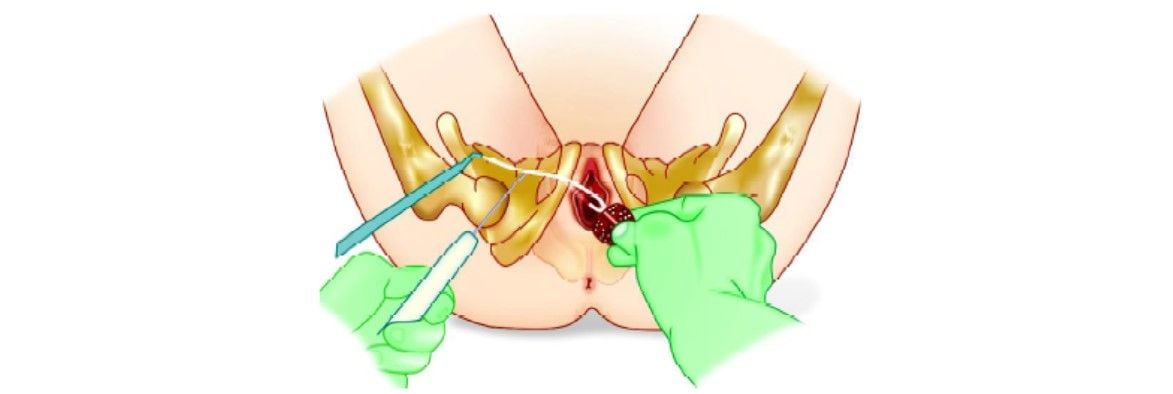
After initiating sharp dissection, continue by using a “push-spread” technique preferably using pointed curved scissors. The path of the lateral dissection should be oriented at a 45° angle from the midline, with the scissors oriented on the horizontal plane. Continue dissection towards the “junction” between the body of the pubic bone and the inferior pubic ramus and perforate the obturator membrane. Winged guides should be inserted into the dissected trace and through the obturator membrane. Insert the TOT device tip with the helical pressure into the dissected tract following the channel of the winged guide. Push the direction of TOT device inward traversing, and perforating the obturator membrane. Once in the TOT needle is in position, the winged guide should be withdrawn.

Dissect to the obturator membrane and perforate obturator membrane

Insert winged guide and helical passer and then remove the winged guide
Once the winged guide has been removed, move the handle of TOT needle towards the midline to a near-vertical position. Then rotate the handle of the helical passer counter clock-wise for the patient's right side and clock-wise for the patient's left side. The point of the helical passer should exit near the previously determined exit points. Slight skin manipulation may be required particularly in obese patients. When the tip of the plastic tube appears and passes at the skin opening, grasp the extreme point of the tip with a clamp and, while stabilizing the tube near the urethra, remove the helical passer by a reverse rotation of the handle. After removing the helical passer the plastic tube should be pulled completely through the skin until the tape appears.

Rotate helical passer while moving the handle into the midline

Facilitate passage of TOT through the skin incision

Grasp tip of the plastic tube then retract helical passer by reverse rotation

Pull plastic tube and tape completely through the skin
The same procedure of TOT mesh insertion should be repeated for the other side keeping in mind that there should not be twisting of mesh in the midline. When the tape is in position, the plastic sheath that covers the tape should be removed. One blunt instrument (e.g. scissors or forceps) should be placed between the urethra and the tape during removal of a plastic sheath, or use other suitable means during sheath removal, to avoid positioning the tape with tension. Following tape adjustment, close the vaginal incisions. Cut the tape ends at the exit points just below the skin on the inner thigh. Close the skin incisions with suture or surgical skin adhesive. A surgeon should remember that “Looser is Better than Tighter”.

Steps of the introduction of the mesh should be repeated on the patient's other side

Adjust the tape, remove the plastic sheath and close incision
The patient should be positioned in dorsal lithotomy with hips hyperflexed with buttock flush to the edge of the table. Make the exit points by tracing a horizontal line at the level of the urethral meatus, and a second line parallel and 2 cm above the first line. Locate the exit points on the second line, 2 cm lateral of the folds of the thigh. Using Allis clamps for traction make a 1 cm midline vaginal incision starting 1 cm proximal to the urethral meatus.

Position the patient, mark thigh exit point and make a vaginal midline incision
After initiating sharp dissection, continue by using a “push-spread” technique preferably using pointed curved scissors. The path of the lateral dissection should be oriented at a 45° angle from the midline, with the scissors oriented on the horizontal plane. Continue dissection towards the “junction” between the body of the pubic bone and the inferior pubic ramus and perforate the obturator membrane. Winged guides should be inserted into the dissected trace and through the obturator membrane. Insert the TOT device tip with the helical pressure into the dissected tract following the channel of the winged guide. Push the direction of TOT device inward traversing, and perforating the obturator membrane. Once in the TOT needle is in position, the winged guide should be withdrawn.

Dissect to the obturator membrane and perforate obturator membrane

Insert winged guide and helical passer and then remove the winged guide
Once the winged guide has been removed, move the handle of TOT needle towards the midline to a near-vertical position. Then rotate the handle of the helical passer counter clock-wise for the patient's right side and clock-wise for the patient's left side. The point of the helical passer should exit near the previously determined exit points. Slight skin manipulation may be required particularly in obese patients. When the tip of the plastic tube appears and passes at the skin opening, grasp the extreme point of the tip with a clamp and, while stabilizing the tube near the urethra, remove the helical passer by a reverse rotation of the handle. After removing the helical passer the plastic tube should be pulled completely through the skin until the tape appears.

Rotate helical passer while moving the handle into the midline

Facilitate passage of TOT through the skin incision

Grasp tip of the plastic tube then retract helical passer by reverse rotation

Pull plastic tube and tape completely through the skin
The same procedure of TOT mesh insertion should be repeated for the other side keeping in mind that there should not be twisting of mesh in the midline. When the tape is in position, the plastic sheath that covers the tape should be removed. One blunt instrument (e.g. scissors or forceps) should be placed between the urethra and the tape during removal of a plastic sheath, or use other suitable means during sheath removal, to avoid positioning the tape with tension. Following tape adjustment, close the vaginal incisions. Cut the tape ends at the exit points just below the skin on the inner thigh. Close the skin incisions with suture or surgical skin adhesive. A surgeon should remember that “Looser is Better than Tighter”.

Steps of the introduction of the mesh should be repeated on the patient's other side

Adjust the tape, remove the plastic sheath and close incision


