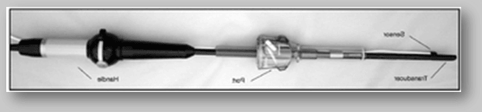
Ultrasonography equipment has two components: a probe and a scanner connected via a cable. The most important characteristics of an operating room ultrasound scanner are compact, mobile, real-time B-mode system and high quality image.1 Doppler capabilities, preferably color Doppler, are highly desirable and, in fact, most important in laparoscopic use to allow visualization of tubular and vascular structures. Further improvements—such as real time 3D visualization by combining several series of 2D images and possible combining of the preoperative computed tomography (CT) scan data with the “live” ultrasound data for improved imaging—are being developed. In the beginning, the available trans-luminal probes were used via laparoscopic ports. The image was 360º with 1-4 cm depth. To allow for good acoustic scanning the probes were enveloped in a sterile plastic sheath filled with saline. These were cumbersome to use and had poor image quality.With the development of dedicated linear array probes, many of these issues have been solved. Ideally, the probe should have a diameter of less than 10 mm to allow introduction through an 11 mm laparoscopic port. The length of the probe should be 35-50 cm to allow access to all locations in the abdominal cavity. Most commonly used linear array probes have frequencies of 5-10 MHz, with depth of penetration of approximately 4-10 cm. A flexible tip will allow for improved scanning angles.
In contrast to the good outcomes of gastric cancer patients reported by Japanese authors, in the United States and rest of the Western world, gastric cancer patients continue to have very poor prognoses. The main factor for this discrepancy seems to be the more advanced stage of disease at presentation for the non-Japanese patients: Up to 28% of patients with gastric cancer are found to have unresectable disease at the time of their surgeries because of local extension or peritoneal metastasis despite negative preoperative imaging studies. The rationale for the use of staging laparoscopy and LUS in patients thought to have resectable disease based on preoperative imaging is to decrease the rate of negative laparotomies and the associated morbidity. Preoperative laparoscopy is recommended by the National Cancer Institute as part of the routine staging of gastric cancer patients. To rule out metastasis disease or local extension, any patient considered to be a surgical candidate should undergo preoperative laparoscopy. If no evidence of metastasis disease is found during laparoscopic inspection, LUS has the potential to increase the yield of the procedure by identifying lymph node involvement and deep liver metastasis and by allowing directed biopsies.
Most commonly, three ports are used. A 10 mm port is placed at the umbilicus. A 5 mm trocar is placed in the left upper quadrant and a 10-12 mm trocar is placed in the right upper quadrant. After a complete laparoscopic examination has been performed and peritoneal lavage obtained for cytology, a flexible tip laparoscopic ultrasound probe is introduced into the abdomen through the umbilical port. The liver is evaluated/fixed first. Both the left and right lobes are evaluated from the anterior and posterior surfaces. Then the celiac axis and hepato-duodenal ligament are examined. Color-coded Doppler flow further helps differentiate blood vessels from lymph nodes. Similarly para-aortic lymph nodes are fixed. Finally, the primary tumor is assessed for local invasion and resectability. Based on the LUS findings, biopsies can be performed or lymph nodes exercised. Generally in Diagnostic laparoscopy (DL) for gastric cancer there is a small risk for conversion to an open procedure and in frequently may lead to bleeding. The addition of laparoscopic ultrasound to this procedure has not been reported to increase this risk. DL with ultrasound upstages gastric cancer patients in up to 40% of cases and prevents laparotomy in roughly 25% of the patients (Level II). Again, the procedure may also help downstage more advanced tumors—from T4, based on endoscopic ultrasound (EUS), to T3—by ruling out direct invasion of surrounding structures. Ultrasound has also been demonstrated to allow the detection of M1 nodes in the hepatoduodenal ligament. In contrast, some studies have reported no benefit of LUS when added to a routine staging laparoscopy (Level 3). All of the studies are retrospective reviews of collected data, but they do show a significant decrease in negative laparotomy rates. The difficulty in quantifying the added benefit of LUS over routine DL is another limitation, though as mentioned above, there seem to be some advantages. One study reports that LUS added additional information (over laparoscopy alone) in 1 out of the 28 patients who had unresectable disease as determined by staging laparoscopy (Level III). LUS for the staging of gastric cancer patients can be performed safely, adds little time to the duration of staging laparoscopy, and does not increase significantly patient morbidity (Grade A). The routine use of staging laparoscopy and LUS after a negative preoperative work-up (CT with or without EUS) is recommended.
Guidelines for the Use of Laparoscopic Ultrasound in Esophagogastric Cancer
Although laparoscopic examination alone identifies most cases of metastasis disease, such as superficial hepatic lesions and peritoneal seeding, the addition of LUS benefits a subset of patients.1-4 No additional morbidity has been reported for LUS when added to staging laparoscopy. Its use increases the operative time by 15-20 minutes, for the added diagnostic benefit it seems reasonable. Laparoscopic ultrasound has been used in adrenal surgery, largely to help localize tumors. Although it is not necessary in most cases, it may add information that is helpful to identify the extent of the disease in the removal of a tumor. Laparoscopic ultrasound can be used to localize a gland, determine the invasion of tumors into adjacent organs, identify adrenal veins/arteries, and identify pathology in other organs. Set-up/arrangement is the same as for any laparoscopic adrenalectomy. The initial camera port must be 12 mm to allow for the size of the flexible ultrasound probe. On the right side, the ultrasound may be performed after retraction of the liver anteriorly and then after dividing the posterior peritoneal attachments of the liver, if necessary, to identify the cephalad portion of the gland. On the left side, identification of the gland is performed after medial rotation of the spleen. On both sides, the gland is identified by its relationship to the kidney. Adenomas are homogeneous in appearance, and pheochromocytomas are heterogeneous.
Complexities are uncommon, but misidentification or mislocation could lead to harm. Benefits are the identification of the adrenal gland when it is difficult to find in the retroperitoneal adipose tissue, the identification of adrenal vasculature, and the assessment of adjacent organs for invasion. Pathology in other organs can also be assessed. LUS can benefit laparoscopic adrenalectomy by localizing the gland, assessing for invasion into adjacent organs, and assessing pathology in other organs 39% of the time. The adrenal vein can be identified with ultrasound 21% of the time. The mean time of use is 10.9 minutes, (Level III).
Guideline for the Use of Laparoscopic Ultrasound in Biliary Disease
An alternative to intraoperative cholangiography to identify choledocholithiasis during laparoscopic cholecystectomy as has been the primary use of LUS in biliary disease. LUS can be used routinely to identify the common bile duct, common hepatic duct, and intra-hepatic bile ducts to determine dilation or bile duct stones. In some cases, the cystic duct anatomy can also be discerned.
Use of general anesthesia is common, and port placement is the same as for laparoscopic cholecystectomy. The placement of probes will be different for patients with hepatomegaly. The flexible ultrasound probe is placed through the umbilical port, while the camera is placed through the midepigastric port. First, the liver is scanned and the common bile duct can be seen medial to the gallbladder. The gallbladder and liver are retracted superiorly and cephalad so that the transducer can be placed directly over the common bile duct. Sometimes the junction of the right and left hepatic ducts or the cystic duct junction can be seen. The common bile duct is followed to the duodenum. A transverse view of the bile duct can be obtained by acute deflection of the transducer. In the duodenum if there is air, it can be compressed with the probe or water can be instilled into the stomach for a better view of that area. A cholangiogram catheter can be placed into the cystic duct and flushed with saline to distend the duct If the common bile duct is hard to visualize. The common bile duct can be fully evaluated in 97% of cases (Level II). For identifying choledocholithiasis, the sensitivity is 90%-96% and specificity 100% (Level II). The positive predictive value is 100%, and the negative predictive value is 98% (Level II). In a comparative trial, intraoperative cholangiography had a sensitivity of 86%, specificity of 99%, positive predictive value of 98%, and negative predictive value of 92% (Level II).
LUS is a good alternative to intraoperative cholangiogram (Grade B). Compared to intraoperative cholangiogram, it costs less to perform (Grade B) and takes less time (Grade C).
Guidelines for the Use of Laparoscopic Ultrasound in Gynecologic Procedures
LUS has not been in wide use in gynaecologic procedures, although the available data suggest it could potentially be a helpful modality for ascertaining lymph node metastases for cervical carcinoma, identifying myomas of the uterus when they are not overtly apparent during laparoscopic surgery, estimating ovarian cysts. Indications for LUS for gynecologic conditions include ovarian masses, uterine masses, particular myomas, and staging in early stage cervical carcinoma.
For uterine or ovarian masses, port set-up position would be the same as for laparoscopic removal with the only exception that one of the ports needs to be 12 mm in size to pass the ultrasound through the port. A flexible ultrasound should be used to be able to evaluate any masses from multiple directions. Saline must be added if there is too much air between the transducer and the object being identified. LUS for identifying lymph node metastases with cervical carcinoma can be performed through a two-port technique: a camera inserted through periumbilical port and a 12 mm port placed supraumbilically; in this case, saline must be used as an acoustic medium/median. The ultrasound probe is placed along the ilia vessels identifying the lymphatic chain up to the renal vessels. The use of Doppler can differentiate blood vessels from solid masses to help direct biopsies. Any lymph node identified as being >1 cm would be exercised using this technique. The LUS procedure is much safer. The main benefit is improved diagnostic accuracy compared to other radiologic methods. For cervical carcinoma, lymph node metastasis sensitivity is 63%-91% and specificity 95%-100%, with an accuracy of 87%, positive predictive value of 82%, and negative predictive value of 89%.All this is an improvement over all other modalities including CT, magnetic resonance imaging, and positron emission tomography scans. Sentinel node mapping has been utilized for cervical carcinoma; however, it is known to miss some metastases. The important problem with ultrasound detection of lymph nodes in cervical cancer is that it misses microscopic disease. Currently, LUS does not obviate the need for pelvic lymph node dissection and is not in widespread use despite its accuracy (Level II).For ovarian cysts, LUS has been used to help with location and for diagnosis. Accurate diagnosis with the use of LUS is 85% whereas transvaginal ultrasound is 64%; however, this is not significantly different in one small series. Laparoscopic ultrasound may identify the location of a cyst within an ovary and therefore decrease damage to the ovary doing decortication. Laparoscopic ultrasound can also be used intra-operatively to identify the location of myomas in the uterus to help with their removal.
Guideline for the Use of Laparoscopic Ultrasound in Kidney Disease
Trans-abdominal ultrasound is a very important modality for identifying kidney disease. LUS is used to assist in identifying structures during operation. LUS has been used in a variety of laparoscopic procedures related to the kidney. These include the drainage of lymphoceles following renal transplantation, cyst marsupialization, and guidance for radiofrequency ablation of renal tumors. The procedure is performed under general anesthesia. The patient is positioned in a lateral decubitus or semi-decubitus position with the diseased kidney up. Port positioning depends on the procedure being performed and the surgeon’s preference; however, the ultrasound port needs to be the right size for the probe. LUS may help decrease the incidence of inadvertent intra-operative injuries (Level III). Nonetheless, a review of 20 studies and 57 patients undergoing laparoscopic drainage of post-transplant lymphoceles revealed a 7% recurrence rate and a 5.3% complication rate, which were slightly higher than the results for open drainage (3.8% and 3.8%, respectively) (Level III). In contrast, the largest single institution series of laparoscopic drainage of post-transplant lymphoceles (n=63 patients) showed a similar recurrence rate to open drainage (7% for both) but a lower rate of complications (6% v/s 11%, respectively) (Level III). Operative time, conversions, length of hospital stay, and complications improved substantially over an 8-year period.
LUS allows lymphoceles to be drained with minimally invasive techniques and early discharge from the hospital. In another series of 19 patients, all but one patient were discharged home within 24 hours after laparoscopic drainage of lymphoceles (Level III). Similar results have been reported for the marsupialization of simple renal cysts and autosomal dominant polycystic kidney cysts with good postprocedure improvement in pain.3,5,6 LUS has been used in these studies for the localization of deep cysts. LUS is invaluable for radiofrequency ablation treatment of renal tumors. Studies document good results up to 1 year after its use (Level III).
Guidelines for the use of Laparoscopic Ultrasound in Liver Disease
The primary role of LUS in liver disease is to assist staging of hepatocellular cancer or metastasis of colon cancer. LUS is indicated for staging hepatocellular cancer and metastatic colon cancer. It has also been used to identify metastasis for pancreatic and gastric cancers. To identify the extent of cystic disease, ensure complete decortication of cysts, and assist with identifying the extent of benign and malignant masses such as hemangiomas prior to laparoscopic resection or ablation, LUS is used. Several techniques could be used for staging purposes, and we will describe a commonly used approach. General anesthesia is typically used. The primary trocar is placed periumbilically with two secondary trocars placed subcostally at the xyhoid or under the left costal margin and between the midaxillary line and the anterior axillary line. Through these ports, all areas of the liver should be viewable. Lesions can be biopsied or ablated under laparoscopic guidance. The extent of lesions and the proximity to vascular or biliary structures can be evaluated. The procedure is terminated if lesions are determined to be unresectable. If the lesions are resectable, then either a laparoscopic or an open procedure is performed.
When LUS is used for other lesions or cysts, trocar placement will depend on where the lesion or lesions are situated.
The risks of the procedure are that of diagnostic laparoscopy, general anesthesia and biopsy, ablation, or resection (bile leak, bleeding, and seeding). The addition of LUS in and of itself does not increase the risk of the procedure, but for staging, false negative results can lead to unnecessary open surgery. The benefits of LUS for staging are the identification of further lesions and the extent of tumors, especially involvement of ducts or vessels, to aid in planning resection. LUS can determine resectability and obviate the need for open exploration when unresectability is determined. LUS can enable biopsy for tissue diagnosis.
DL, with the addition of LUS for colon and rectal metastasis or hepatocellular cancer staging, identifies 10%-25% more additional tumors than preoperative CT (Level II-III). However, DL can be restricted in 13% of cases and not possible in 3% due to adhesions (Level II). DL with LUS changes the management in up to 49% of cases, and LUS alone added additional staging information in 42% of cases (Level II).5 DL with LUS has specificity of 75%-90% and sensitivity of 80%-100%, with a positive predictive value of 85% (Level III). Unnecessary open surgery for missed disease was uncommon, and avoidance of open surgery due to unresectability was 16%-25% (Level II-III). In pancreatic cancer, DL with LUS was 100% sensitive and specific (Level III).
LUS has been used successfully for liver biopsies in patients with cirrhosis, for decortication of liver cysts, and to determine lines of resection in benign disease (Level III).
Guidelines for the Use of Laparoscopic Ultrasound for the Pancreas
Pancreatic adenocarcinoma has a dismal prognosis, with <5% overall 5-year survival. Despite the availability of high quality preoperative imaging studies, 11%-48% of patients are found to have unresectable disease at the time of laparotomy. To minimize the number of unnecessary laparotomies, staging laparoscopy has been introduced in the treatment algorithm of pancreatic adenocarcinoma patients. Nevertheless, locally advanced or metastatic disease is missed by staging laparoscopy in 5%-25% of patients due to the inability to evaluate vascular invasion and deep hepatic metastases. LUS enables the detection of vascular invasion and deep hepatic metastases and, therefore, has the potential to decrease the false negative rate of staging laparoscopy. In addition, LUS can also have merit in the detection of benign pancreatic diseases.
By identifying occult metastatic disease to the liver or unsuspected locally advanced disease not seen on preoperative imaging the procedure is indicated for the enhancement of the diagnostic accuracy of staging laparoscopy. It can also be used for the localization of nonpalpable and visually occult malignant or benign lesions of the pancreas and to guide pancreatic procedures such as enucleation of insulinomas. The feasibility of LUS for pancreatic adenocarcinoma has been demonstrated in multiple studies (with success rates ranging from 94% to 100% (Level II, III). The main cause of technical failures are dense adhesions that impair examination with the ultrasound probe. Nevertheless, even patients with adhesions can be examined; however, the extent and yield of the examination may be compromised. Conversions to open surgery are uncommon and have been reported in <2% of patients in a large series study (Level III). The procedure is usually performed under general anesthesia, and the majority of reports have used 15 mmHg insufflation pressures. A detailed LUS examination is employed during which the deep hepatic parenchyma, portal vein, mesenteric vessels, celiac trunk, hepatic artery, the entire pancreas, and even pathologic periportal and para-aortic nodes can be evaluated and biopsied ,If no metastatic disease is identified after a thorough visual inspection of peritoneal surfaces. Color-flow Doppler can further assist in assessing the report.
Debate continues about whether LUS should be a component of staging laparoscopy for pancreatic adenocarcinoma patients. Advocates of a short duration procedure that is based only on the inspection of abdominal organ surfaces argue that the procedure can be performed quickly (usually within 10 minutes), can be conducted through one port, does not require significant expertise, poses little risk of potential complications from the dissection near vascular structures, and has good diagnostic accuracy (Level III). On the other hand, advocates for the use of LUS argue that the diagnostic accuracy of staging laparoscopy for pancreatic adenocarcinoma can be enhanced by detecting vascular invasion of the tumor or deep hepatic metastasis, often missed by visual inspection alone, and that it can be performed safely without a significant increase in morbidity and within a reasonable time (Level II, III).
In general more beneficial than complications, the benefits of the procedure include the avoidance of unnecessary exploratory laparotomy, with its associated higher morbidity and cost in patients with metastatic pancreatic adenocarcinoma. Moreover, the procedure can contribute to the selection of more appropriate treatment for patients with true locally advanced disease. On the other hand, false negative studies may lead to unnecessary exploratory laparotomies and unnecessary cost. The procedure affords the patient the advantages of minimally invasive surgery by enabling the detection of deep pancreatic lesions and by guiding laparoscopic surgery. Procedure-related complications are possible but very uncommon. Number of studies have reported on the diagnostic accuracy of laparoscopy with LUS for the staging of pancreatic adenocarcinoma patients, but many do not report separately the added benefit of LUS. In studies reporting the added benefit, LUS detected unresectable disease in 11%-28% of patients whose disease was missed by DL alone, with false negative findings in 1%-8% of patients. It prevents unnecessary laparotomy in up to 34% of patients (Level II-III).7-15 Overall, the reported diagnostic accuracy for DL/LUS ranges between 87% and 100% (Level II-III). Studies comparing DL/LUS with other modalities, such as ultrasound, CT, or EUS, have demonstrated superior results for DL/LUS. Catheline et al17 reported that the sensitivity of DL with LUS was better for the detection of liver metastases (100%), peritoneal metastases (100%), vessel encasement (100%), and nodal involvement (88%) compared to ultrasound, CT, and EUS but less effective for the assessment of the primary tumor (90%; especially for small tumors <2 cm) compared to EUS (100%) (Level II).
The use of LUS for other pancreatic diseases and procedures has been reported to provide useful information for the diagnosis, location, and guidance of the procedure in 60% of patients and to change the surgical strategy in 30%. In addition, reports show intraoperative ultrasound (IOUS) to be useful for detecting and localizing insulinomas, with an accuracy of 83%-100% (Level II-III).This sensitivity has been reported to be stronger than that of preoperative angiography. Recent publications also confirm the ability of LUS to detect neuroendocrine tumors of the pancreas as small as 3-5 mm in diameter.The ability of intraoperative ultrasound to detect extrahepatic gastrinoma is lower (about 58%) (Level III).
That the diagnostic yield of the procedure is highly dependent on operator ability, has a steep learning curve, and depends on the histology, stage of disease, tumor size, and location (Level III).There is convincing evidence that the yield of staging laparoscopy/LUS is significantly higher in patients with pancreatic cancer compared to other types of periampullary tumors (Level III). In addition, the procedure has a higher yield in patients with locally advanced cancer (Level III) and in those with larger tumors (Level III) or tumors located in the pancreas body and tail (Level III). DL/LUS-related morbidity is low and has been reported to range between 0% and 4% (Level II, III). Most complications are minor and consist of wound infections, bleeding at port sites, or skin emphysema. There are no available studies that compare complications between a short-duration procedure only with inspection (DL) and a more extended procedure that combines DL and LUS. Hospital length of stay after staging laparoscopy has been reported to as 1-4 days. Evidence suggests that the hospital stay is shorter after laparoscopic staging compared to open staging in pancreatic cancer patients (Level III).
For more information:
World Laparoscopy Hospital, Cyber City, DLF Phase II, Gurugram, NCR Delhi, 122 002, India
PHONES:
For Training: +919811416838
For Treatment: +919811912768
For General Enquiry: +91(0)124 - 2351555
Email: contact@laparoscopyhospital.com


