The relative position of the instrument ports is very important in the performance of surgical procedures endoscopically. The angle the instruments make with the operative site and to each other should mimic, as far as possible, the natural relationship of the hands and eyes during conventional surgery.
A satisfactory relationship includes:
- An angle of 60 degree between the two instruments tips
- Tangential approach to the site
- Appropriate working distance
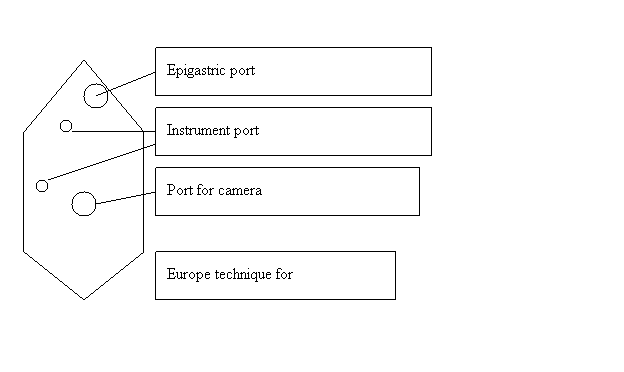
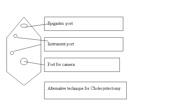
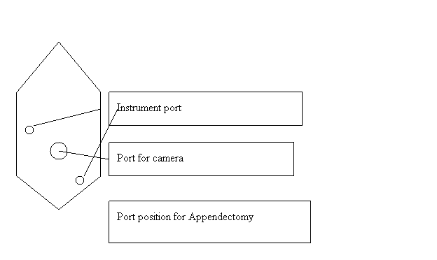
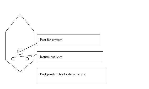
These factors combined with the specific anatomy will determine individual port sites. For standard operations like Cholecystectomy, standard port sites related to surface marking may suffice but as more advanced or varied situations are tackled we recommend that you master the skill of individual port placement using the internal view. In general, the optic and the two main operating ports usually lie at the points of a flattened triangle, the optic being centrally and more distally placed. Try to keep ports at least 5cms apart. (Refer to diagram on next page)
Task analysis and good ergonomic principles are backbone of a laparoscopic surgeon
Port position for Cholecystectomy
Port position for Appendectomy
Port position for bilateral hernia
Swording
Swording occurs when the telescope or the shaft of the assistant’s instrument obstruct the operator’s instruments. If this occurs you may need to consider:
- Repositioning retracting instruments
- Rotation of an angled telescope allowing alteration of the position of the end of the telescope
- Withdrawal of the telescope
- Transposition of the operator’s instruments
- Additional port placement
- changing the instruments to a different port
Sudden collapse
The anesthetist will be checking for conditions such as:
Drug reactions Pneumothorax, Gas embolism myocardial arrhythmia’s
Cardiac arrhythmia’s
- Stop insufflation
- Correct hypoxia and resuscitate
- Postpone surgery
If no other cause of arrhythmias is present assume bleeding to be the cause.
Severe hypotension
Proceed to immediate laparotomy with all instruments left in situ.
Mild to moderate hypotension
The surgeon should consider:
- Discontinuing gas insufflation immediately and reducing intra abdominal pressure to 8.0 mm Hg.
- Proceed immediately to 360° scan of the abdominal cavity for retroperitoneal bleeding.
If bleeding or expanding haematoma is seen, proceed immediately to long midline laparotomy and compression of the bleeding vessel. Aspirate blood, expose and control with vascular clamps. When necessary obtain assistance of a vascular surgeon.
Diagnostic laparoscopy
The usual site of insertion of the trocar/cannula for diagnostic laparoscopy is below or to the side of the umbilicus. This position may require to be altered in the presence of abdominal scars. The use of a 30 degree forward oblique telescope is preferable for viewing the surface architecture of organs. By rotation of the telescope, different angles of inspection can be achieved.
The first important step after access to the abdomen has been gained is to check for damage caused by trocar insertion. A second 5 mm port may then be inserted under vision in an appropriate quadrant to take a palpating rod.
A systematic examination of the abdomen must then be performed just as in laparotomy. We begin at the left lobe of the liver but any scheme can be used as long as it is consistent. Next check around the falciform ligament to the right lobe of liver, gallbladder and hiatus. After checking the stomach, move on to the caecum and appendix and check the terminal ileum. Follow the colon round to the sigmoid colon, and then check the pelvis. You should be conversant with sampling and biopsy techniques, and the use of position and manipulation to aid vision. This is the first procedure to be mastered when learning laparoscopic surgery.
When performing a diagnostic laparoscopy to confirm appendicitis and as a prelude to laparoscopic appendicectomy, a five mm port is placed in the left iliac fossa to faciliate manipulation. The patient is placed head down and rotated to the left to displace the small bowel from the pelvis and allow the uterus and ovaries to be checked. This however should be limited to avoid contamination of subphrenic spaces if this is not already present.


