
Page 45 - World Journal of Laparoscopic Surgery
P. 45
Dysphagia after Bougie-guided Crural Repair
Preoperative Evaluation
Preoperative assessment of the patients was done by a detailed
history taking and physical examination. A 24-hour PH monitoring,
upper gastrointestinal (GI) endoscopy, and esophageal manometry
were done for all patients to reach the exact diagnosis of refractory
GERD, to exclude esophageal motility disorders or achalasia and also
to detect any esophagitis or Barrett’s esophagus caused by GERD.
As posted by DeMeester et al., pathologic reflux was defined
11
by esophageal acid exposure with a DeMeester score greater than
1
14 without having any proton pump inhibitors (PPIs). However,
amplitudes of 30 mm Hg of mean distal esophageal contraction and
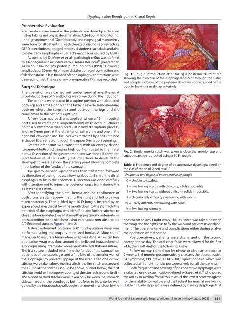
failed peristalsis in less than half of the esophageal contractions were Fig. 1: Bougie introduction after taking a posterior crural stitch
deemed normal. The use of any pre-operative PPIs was recorded. showing the direction of the esophageal descent through the hiatus
and complete closure of the posterior defect was done guided by the
Surgical Technique bougie, leaving a small gap anteriorly
The operation was carried out under general anesthesia. A
prophylactic dose of IV antibiotics was given during the induction.
The patients were placed in a supine position with abducted
both legs and arms along with the table in reverse Trendelenburg
position where the surgeon stood between the legs and the
cameraman to the patient’s right side.
A five-trocar approach was applied, where a 12-mm optical
port (used to create pneumoperitoneum) was placed in Palmer’s
point. A 5-mm trocar was placed just below the xiphoid process;
another 5-mm port at the left anterior axillary line and one in the
right mid-clavicular line. The liver was retracted by a self-retained
S-shaped liver retractor through the upper 5-mm port opening.
Greater omentum was transected with an energy device
(Ligasure–Medtronic) starting high up 4-cm distal to the hiatal Fig. 2: Single anterior stitch was taken to close the anterior gap and
hernia. Dissection of the greater omentum was done till complete smooth passage is checked using a 50 Fr bougie
identification of left crus with great importance to divide all the
short gastric vessels above the starting point allowing complete
mobilization of the fundus of the stomach. Table 1: Frequency and degree of postoperative dysphagia based on
12
the classification of Saeed et al.
The gastro–hepatic ligament was then transected followed
by dissection of the right crus, allowing about 2–3 cm of the distal Frequency and degree of postoperative dysphagia
esophagus to be in the abdomen. Dissection was done carefully 0 = Unable to swallow.
with attention not to injure the posterior vagus trunk during the I = Swallowing liquids with difficulty, solids impossible.
posterior dissection.
After identifying the hiatal hernia and the confluence of II = Swallowing liquids without difficulty, solids impossible.
both crura, a stitch approximating the right and left crus was III = Occasionally difficulty swallowing with solids.
taken posteriorly. Then guided by a 50 Fr bougie, inserted by an IV = Rarely difficulty swallowing with solids.
experienced anesthetist from the mouth down to the stomach, the
direction of the esophagus was identified and further stitches to V = Swallowing normally.
close the hernial defect were taken either posteriorly, anteriorly, or
both according to the hiatal size using interrupted non-absorbable anesthetist to avoid tight wrap. The last stitch was taken between
2/0 Ethibond sutures Figures 1 and 2. the wrap and the right crus to fix the wrap and prevent its displace-
A short redundant posterior 360° fundoplication wrap was ment. The operative time and complications either during or after
performed using the properly mobilized fundus. A “shoe-shine” the operation were recorded.
maneuver to ensure a tension-free wrap was done. A 1–2 cm fun- Postoperatively, patients were discharged on the second
doplication wrap was done around the delivered intraabdominal postoperative day. The oral clear fluids were allowed for the first
esophagus using interrupted non-absorbable 2/0 Ethibond sutures. 24 h, then soft diet for the following 7 days.
The first suture included bites from the fundus of the stomach on Follow-up was carried out by phone or clinic attendance at
both sides of the esophagus and a fine bite of the anterior wall of 2 weeks, 1–6 months postoperatively to assess the postoperative
the esophagus to prevent slippage of the wrap. Then one or two GI symptoms, PPI intake, GERD–HRQL questionnaire which was
stitches were taken above the first stitch (the first stitch was around collected at 1 and 6 months postoperatively for all the patients.
the GEJ so all the stitches should be above, but not below, the first Both frequency and severity of postoperative dysphagia were
12
stitch to avoid an improper wrapping of the stomach around itself). evaluated using a classification defined by Saeed et al. who scored
The second or third stitches were taken only between the stomach the ability to swallow from 0 to 5 in which the lowest score was given
sidewall around the esophagus but not fixed to its anterior wall for the inability to swallow and the highest for normal swallowing
guided by the intraesophageal bougie that moved in and out by the (Table 1). Early dysphagia was defined by having dysphagia that
World Journal of Laparoscopic Surgery, Volume 15 Issue 2 (May–August 2022) 141