
Page 4 - World's Most Popular Laparoscopic Journal
P. 4
Khairi Hajaji et al
postoperative period was uneventful and the patient was was not helpful because of unfamiliarity of our radiology
discharged fit on the fourth postoperative day after the full department staff with such conditions.
course of anthelminthic therapy (mebendazole) for three Anthelminthic therapy with mebendazole or albendazole
days. Patient was seen in surgical clinic and doing well. is a part of conservative management, reported by some
authors with a success rate upto 80% of patients and
DISCUSSION considered as the first line of treatment in the first few
days. 13,14 The rational for initial administration of
Ascariasis is a helminthic infection of global distribution
with more than 1.4 billion persons infected throughout the anthelminthics is to paralyze the parasites within the intestinal
3
world. The majority of infections occur in the developing lumen then worms are expelled by normal gastrointestinal
6,8
countries of Asia and Latin America. It is estimated that peristalsis. However, it is not advisable to have dead
around 20,000 deaths occur per year because of severe worms inside the ductal system, which might lead to stricture
clinical disease caused by ascariasis. 4,5 formation as a result of severe inflammatory reaction. In
Khuroo et al reported 500 cases in India of hepatobiliary addition, the liberated ova or existence of fragmented Ascaris
1,15
and pancreatic diseases due to Ascaris lumbricoides from might act as a nidus for stone formation. Therefore, for
one center over the period from June 1983 to November the above-mentioned reasons and in failed medical treatment
1989. Since then hepatobiliary and pancreatic ascariasis of confirmed biliary ascariasis, endoscopic intervention is
(HPA) has been reported more often than ever before from indicated.
6
many centers in endemic areas. Another 300 cases of HPA ERCP has an advantage as a diagnostic tool as well as
7
were reported in Syria by Sandouk et al. Ascaris causes therapeutic modality. It allows better identification of worms
pancreatitis due to obstruction of papilla of Vater, invasion in the duodenum and those across the papilla, and can be
of common bile duct, or invasion of pancreatic duct and used for worm extraction from the ampullary orifice, biliary
7,16
can occur with abdominal pain, back pain, emesis, fever, duct or pancreatic duct in 98% of patients. Most worms
8
or jaundice. However, the disease is now encountered with were extracted by flushing the bile ducts, grasping forceps,
increased frequency in the western countries. 9,10 or balloon catheters. Thus, ERCP has now become the
The diagnosis of ascaris pancreatitis requires a high investigation modality of choice. Surgical intervention with
degree of suspicion in population at risk. Ultrasonography worm extraction from CBD combined with cholecystectomy
is a simple, noninvasive test and the characteristic sono- should be advised for patients for whom conservative and
graphic findings of worms in the ducts have been well endoscopic management has failed or complicated by
13
described. 8,11,12 The worms move freely in and out of the cholangitis.
biliary tree and ultrasonography cannot diagnose ascariasis Until recently, the conventional open method is the
in the duodenum, therefore more than half of the patients standard surgical treatment for biliary ascariasis involving a
6,7
with HPA would be missed. In our case, the ultrasound combination of cholecystectomy, extraction of parasites,
and T-tube drainage. 6,13 Yoshihara S et al reported the first
case in 2000 of a laparoscopic extraction of living worm
from CBD through a conventional choledochotomy with
primary suture of CBD opening without using drain. 17
18
Astudillo AJ et al had a series of 13 patients diagnosed
with biliary ascariasis diseases between February 1992 and
February 2007. Six of those patients needed laparoscopic
cholecystectomy and extraction of worms from CBD with
insertion of T-tube, only one patient had primary closure of
CBD. There is another reported case of laparoscopic
extraction of worm without the need for T-tube drainage
reported by Moirangthem GS et al. 19
In our case, this is a first reported case of living worm
being extracted laparoscopically through a cystic duct
opening using intraoperative cholangiography with a few
millimeters extension to the junction between cystic duct
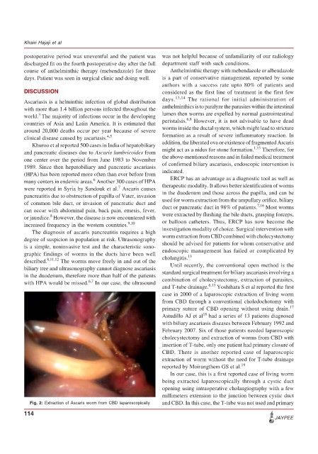
Fig. 2: Extraction of Ascaris worm from CBD laparoscopically and CBD. In this case, the T-tube was not used and primary
114
JAYPEE