
Page 10 - WJOLS - Laparoscopic Journal
P. 10
Falih Mohssen Ali
age 43.4 years). The most common complaints were dull laparoscope was inserted into the cyst to exclude any biliary
pain at the right hypochondrium or/and epigastrium and communication or retained daughter cysts. The cystic cavity
palpable mass. Patients were diagnosed by ultrasonography was irrigated with 20% hypertonic saline several times, and
(US) (Fig. 1), computed tomography (CT), magnetic partial or near total cystectomy was done by using harmonic
resonance imaging (MRI) and confirmed by serological scalpel, then a drain was placed in the remaining cystic
examination (immunoelectrophoresis which has a high cavity, and gauzes were placed in an endosac and removed.
sensitivity, being positive in 30 patients). We excluded cases Postoperative follow-up was very smooth, oral fluid intake
with multiple liver hydatid cysts having more than two or was allowed next day of operation, drain was removed
cysts located in blind area for laparoscopic procedures, like at 48 hours after operation if no apparent bile in the drain,
segments 1, 2 and 7. Our exclusion criteria also included patients were discharged to home and advised for follow-up
intraparenchymal location of the cyst or cysts with thick at 2 weeks, 3 months and 6 months and then yearly by
and calcified walls. All procedures were performed under ultrasound and serological tests (immunoelectrophoresis
general anesthesia and in the supine position. Prophylactic test). 5,8,11
antibiotics were administered for 30 minutes before the
operation. The surgeon and the camera assistant standing Indications
on the left side of the patient with the assistant and scrub 1. Single superficial cyst that may rupture
nurse standing on the right side of the patient. Four ports 2. Large cyst with multiple daughter cysts
were placed, a supraumbilical 10 mm port through which a 3. Cysts in communication with the biliary tree
0° telescope inserted, another 10 mm port inserted at the 4. Infected cysts
epigastrium as near as possible to the cyst and used as a 5. Cysts giving compression to the near vital organs.
working channel and two additional 5 mm ports inserted
4
according to the cyst location. From the epigastric port, Contraindications
gauzes soaked with 20% hypertonic saline as scolicidal agent
were introduced into the abdominal cavity and placed around 1. Dead cysts
the cyst. The cyst was punctured with long laparoscopic 2. Multiple cysts
needles connected to vacuum suction through epigastric 3. Cysts difficult to access
port; another sucker was introduced through the right 4. Small cysts.
5 mm port to avoid accidental spillage of the cyst content.
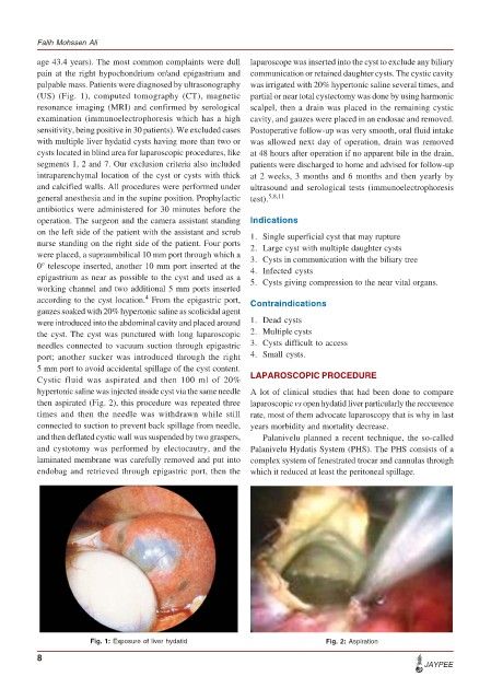
Cystic fluid was aspirated and then 100 ml of 20% LAPAROSCOPIC PROCEDURE
hypertonic saline was injected inside cyst via the same needle A lot of clinical studies that had been done to compare
then aspirated (Fig. 2), this procedure was repeated three laparoscopic vs open hydatid liver particularly the reccurence
times and then the needle was withdrawn while still rate, most of them advocate laparoscopy that is why in last
connected to suction to prevent back spillage from needle, years morbidity and mortality decrease.
and then deflated cystic wall was suspended by two graspers, Palanivelu planned a recent technique, the so-called
and cystotomy was performed by electocautry, and the Palanivelu Hydatis System (PHS). The PHS consists of a
laminated membrane was carefully removed and put into complex system of fenestrated trocar and cannulas through
endobag and retrieved through epigastric port, then the which it reduced at least the peritoneal spillage.
Fig. 1: Exposure of liver hydatid Fig. 2: Aspiration
8
JAYPEE