
Page 24 - World Journal of Laparoscopic Surgery
P. 24
Mini Two-port Laparoscopic Appendicectomy with Novel Knotting Technique
port. Young cosmesis oriented patients with acute appendicitis and then the tip of appendix is then repositioned within 2-0
without lump or perforation, recurrent appendicitis having polypropylene loop. Mesoappendix is divided with bipolar energy
symptoms due to fecolith, and incidental finding of inflamed device till base is visible (Fig. 1). A segment of 2-0 polyglactin suture
appendix in diagnostic laparoscopy. Preileal, subceacal, and pelvic held on tip of needle holder together is introduced through the
position of appendix were preferred. 5-mm suprapubic port so as to encircle the base of the appendix.
After encircling the base and creating a loop, tip of the 2-0
operAtIve technIque polyglactin suture is again held with needle holder in the right
Under general anesthesia, patient is placed in Trendelenburg hand of surgeon and with outer end of 2-0 polyglactin suture held
in surgeon’s left hand, and single instrument surgical knot analogs
position with laparoscopy trolley on patient’s right and surgeon to the open technique is performed (Fig. 2), wherein internal end
on patient’s left side. Laparoscopic access into the abdomen of the suture is held with needle holder in the right hand and the
was obtained via Hasson’s technique through the umbilicus long end of 2-0 polyglactin suture is held externally by the left hand.
with 5-mm port, and the procedure was started by creating Another knot is placed at the distal location in the similar fashion
pneumoperitoneum through umbilical port with insufflation and appendix is divided and delivered through either of 5-mm
pressures being maintained between 10 and 12 mm Hg. A 5-mm 30° port after completely withdrawing specimen within the cannula
scope is introduced through the 5-mm umbilical port. Under direct of 5-mm port to prevent port-site contamination.
vision, a 5-mm trocar was inserted through a suprapubic incision
made below the pubic hairline (Fig. 1). A 2-0 polypropylene suture In a Case of Grossly Inflamed Appendix/Edematous
is threaded and reversed through an 18-gauge epidural needle to Cecum
create a loop at the tip. This needle loop retractor is then inserted In an instance of the edematous cecum and grossly inflamed
in the right iliac fossa (Fig. 1) at the position of appendix as defined appendix, base of the appendix is transfixed (Fig. 3) with entire
by laparoscopy. Dissection of mesoappendix up to the base of the length of 2-0 polyglactin suture introduced through a percutaneous
mesoappendix is done using bipolar energy device (Fig. 1).
puncture in right iliac fossa, needle is cut and retrieved through right
In Case of Dense Adhesions and When Tip of Appendix iliac fossa, and opposite long end of suture is pulled out through
is Not Visualized 5-mm port alongside of the needle holder. Knot analogous to the
In difficult appendix with adhesions and when the tip of the open surgical knot is placed as described above, and then second
surgical intracorporeal knot is placed distally. Appendix is divided
appendix is not visualized, a double loop retraction, one with between two knots and retrieved. None of the operated cases
additional subserosal appendix stitch with 2-0 polyglactin suture were converted to conventional 3-port or open appendicectomy.
passed through abdominal wall is taken on most visible portion of
appendix which aides in retraction and dissection of the appendix,
and when tip becomes visible, a second 2-0 polypropylene loop results
retraction as described above is used (Fig. 1) to hitch up the A total of 200 patients were operated of which 168 underwent TPA
appendix and aid in the process of adhesiolysis (Fig. 1). After and 32 underwent CLA. Comparison of the two group’s operative
adequate mobilization, first polyglactin suture is later removed time was 24 minutes and 42 minutes for TPA and CLA, respectively.
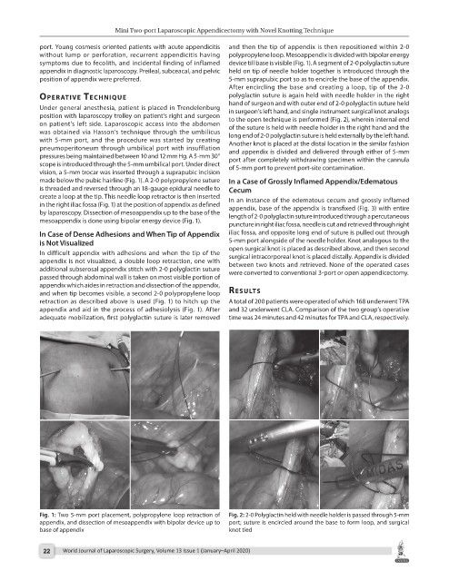
Fig. 1: Two 5-mm port placement, polypropylene loop retraction of Fig. 2: 2-0 Polyglactin held with needle holder is passed through 5-mm
appendix, and dissection of mesoappendix with bipolar device up to port; suture is encircled around the base to form loop, and surgical
base of appendix knot tied
22 World Journal of Laparoscopic Surgery, Volume 13 Issue 1 (January–April 2020)