
Page 40 - World Journal of Laparoscopic Surgery
P. 40
Anshika Lekhi et al
Fig. 2: Uterine horn Fig. 3: Bicornuate uterus
Fig. 4: Uterine septum Fig. 5: Hysterosalpingograph
revealed few adhesions for which adhesiolysis was
done and the cavity was normalized. She was ad-
vised for normal trial of conception. Her intrauterine
pregnancy was confirmed at 6 weeks and 4 days after
6 months of surgery.
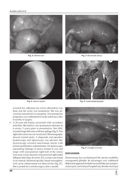
6. A 26-year-old female presented with secondary
infertility. She had h/o one spontaneous abortion at
6 weeks, 3 years prior to presentation. Her HSG
revealed single left cornu with free spillage (Fig. 5). The
right-side cornu was not visualized. Ultrasonography
showed normal study. A diagnostic and operative
hysteroscopy and laparoscopy was advised. Her
hysteroscopy revealed unicornuate uterus with
normal proliferative endometrium. On laparoscopy,
astonishing findings of uterus normal in size and Fig. 6: Complex anomaly
shape with noncanalized right half of the uterus DISCUSSION
were seen; it also showed a noncanalized right-sided
fallopian tube (Figs 1K and 6). B/L ovaries were found Hysteroscopy has revolutionized the uterine morbidity
to be normal. Hysteroscopically, lateral metroplasty management globally. Its advantages over traditional
with cavity enhancement was done for her (Fig. 1L). abdominal approach include less morbidity, less postpro-
She is posted for a relook surgery after a month. cedure pain, and reduced hospital stay, thereby making it
88